Microscopic characterisation of Ni nanoparticles exsolution in La0.4CaxSr0.4-xNi0.06Ti0.94O3-d perovskite oxides
- Abstract number
- 151
- Presentation Form
- Poster
- DOI
- 10.22443/rms.mmc2023.151
- Corresponding Email
- [email protected]
- Session
- Poster Session One
- Authors
- Ms SeoJin Kim (1), Dr David N. Miller (1), Prof John T.S. Irvine (1)
- Affiliations
-
1. University of St Andrews
- Keywords
Perovskite Exsolution SEM EBSD STEM
- Abstract text
Perovskite oxides are known to exsolve metal nanoparticles at elevated temperatures in a reducing atmosphere. This phenomenon has been suggested as an alternative process for designing metal nanoparticles to the conventional deposition method because of the enhanced stability and better resistance against agglomeration.1-3 This has been demonstrated in electrochemical applications including solid oxide cells.4 Understanding the mechanisms behind this process will enable optimisation of the properties. A combined SEM, EBSD, and probe corrected STEM study has been carried out to understand how substituting Ca for Sr on the perovskite A-site influences the exsolution of Ni from the perovskite B-site.
A series of La0.4CaxSr0.4-xNi0.06Ti0.94O3-d where x = 0, 0.2, and 0.4 were synthesized by solid-state synthesis. To exsolve nanoparticles, polished pellets were reduced in a 5% H2/N2 atmosphere at 900°C for 10 hours. Thermogravimetric analysis was performed to determine the extent of reduction in each sample. SEM images were taken to observe the size and morphology of exsolved nanoparticles. Electron Backscatter Diffraction was used to determine the effect of perovskite orientation on particle size and morphology, and to allow the preparation of TEM samples from specific crystallographic orientations. Probe corrected STEM imaging was used to characterise the nanoparticle-perovskite interface.
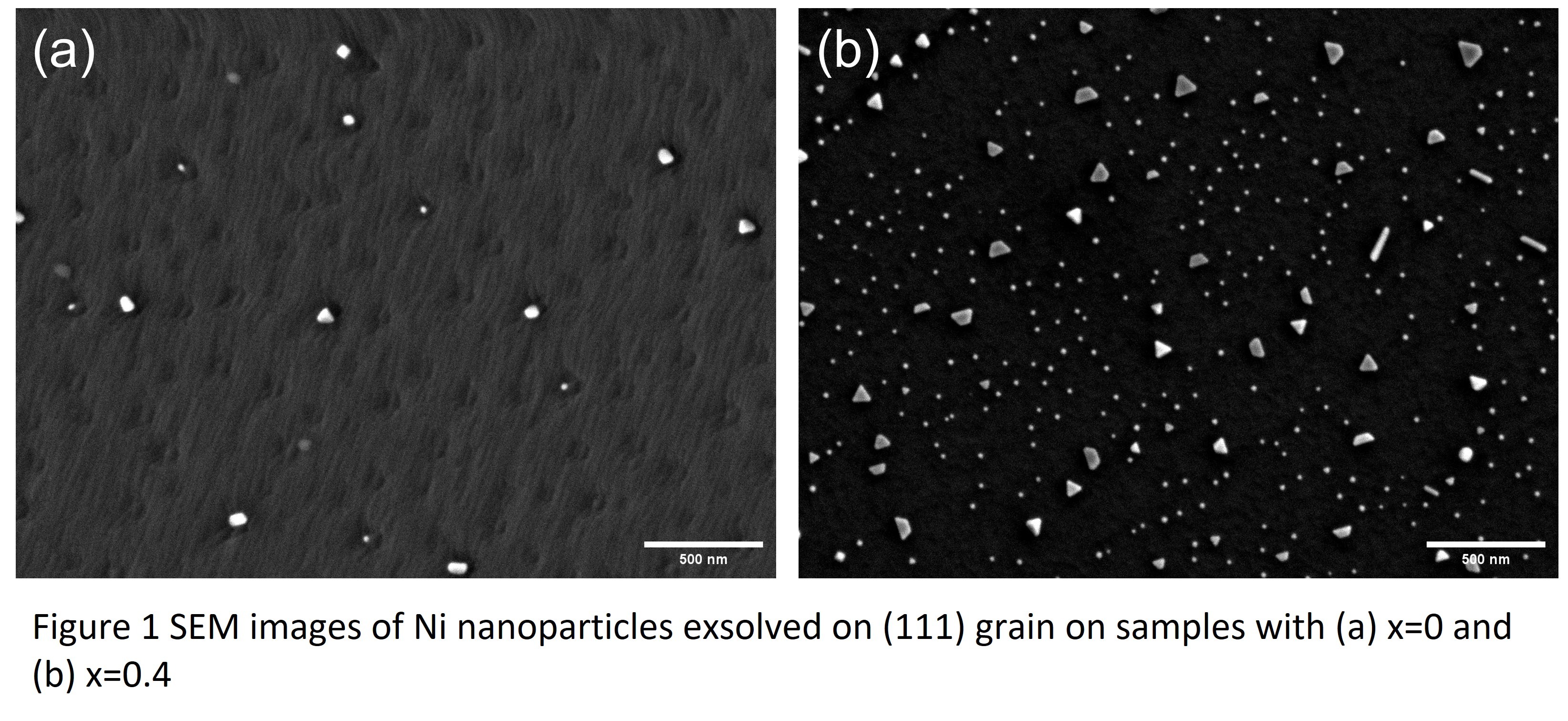
SEM imaging and EBSD analysis have demonstrated that the level of Ca substitution as well as the perovskite orientation has a strong influence on particle populations, size, and morphology. The SEM images of reduced x = 0 and x = 0.4 (Fig. 1) show a greater particle population on a (100) surface when Sr is replaced by Ca. Also, Ni nanoparticles with various morphologies including spheres, triangles, rods, and trapezoids were observable in x = 0.4 samples. Corresponding STEM images, of these samples, show that these differences correspond to changes in the nature of the nanoparticle-perovskite interface.
- References
1. D. Neagu, G. Tsekouras, D. N. Miller, H. Menard and J. T. Irvine, Nat Chem, 2013, 5, 916-923.
2. D. Neagu, V. Kyriakou, I. L. Roiban, M. Aouine, C. Tang, A. Caravaca, K. Kousi, I. Schreur-Piet, I. S. Metcalfe, P. Vernoux, M. C. M. van de Sanden and M. N. Tsampas, ACS Nano, 2019, 13, 12996-13005.
3. O. Kwon, S. Joo, S. Choi, S. Sengodan and G. Kim, J Phys-Energy, 2020, 2.
4. Q. A. Islam, S. Paydar, N. Akbar, B. Zhu and Y. Wu, Journal of Power Sources, 2021, 492.