MerlinEM, Hybrid Pixel Array Counting Detector for Transmission Electron Microscopy.
- Abstract number
- 210
- Presentation Form
- Poster
- Corresponding Email
- [email protected]
- Session
- Poster Session One
- Authors
- Dr. Matus Krajnak (1), Dr. Adriana L. Klyszejko (1)
- Affiliations
-
1. Quantum Detectors Ltd, R103, RAL, OX11 0QX,
- Keywords
MerlinEM; Merlin 4S; Merlin 4R; Merlin 1R; 4D STEM; ptychography; electron diffraction; EELS
- Abstract text
At the University of Glasgow, experiments with hybrid pixel array counting detectors started with a Medipix2 detector [1]. It was clear that direct electron detection and hardware-based electron counting can offer advantageous imaging capabilities. Subsequently, a Medipix3 detector with a Merlin readout system was commercialised as a MerlinEM detector by a collaboration between the University of Glasgow and Quantum Detectors Ltd. With more than 60 systems worldwide, the detector has been applied in multiple experimental configurations.
The detector has been mainly applied in scanning transmission electron microscopy (STEM), however, diffraction and EELS applications are also wide-ranging. [2]
In STEM, the ability to collect a full distribution of electrons for each probe position with a millisecond and better timescales can be readily used in DPC [3] and ptychography [4] but it also enables a more quantitative approach to standard techniques and potential for development of new ones.
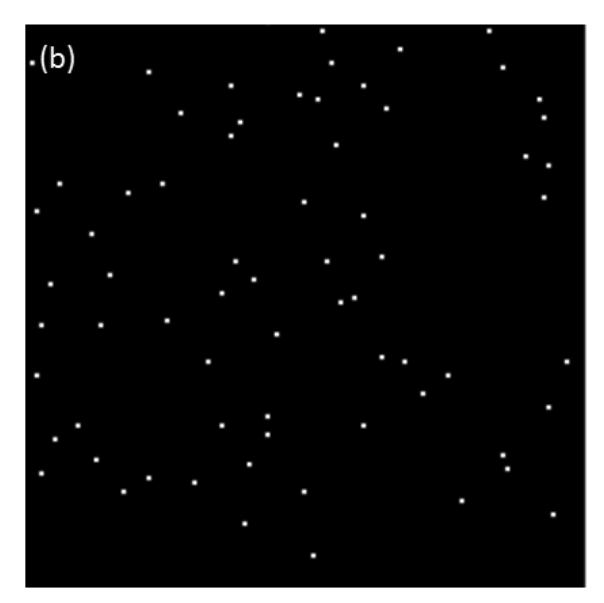
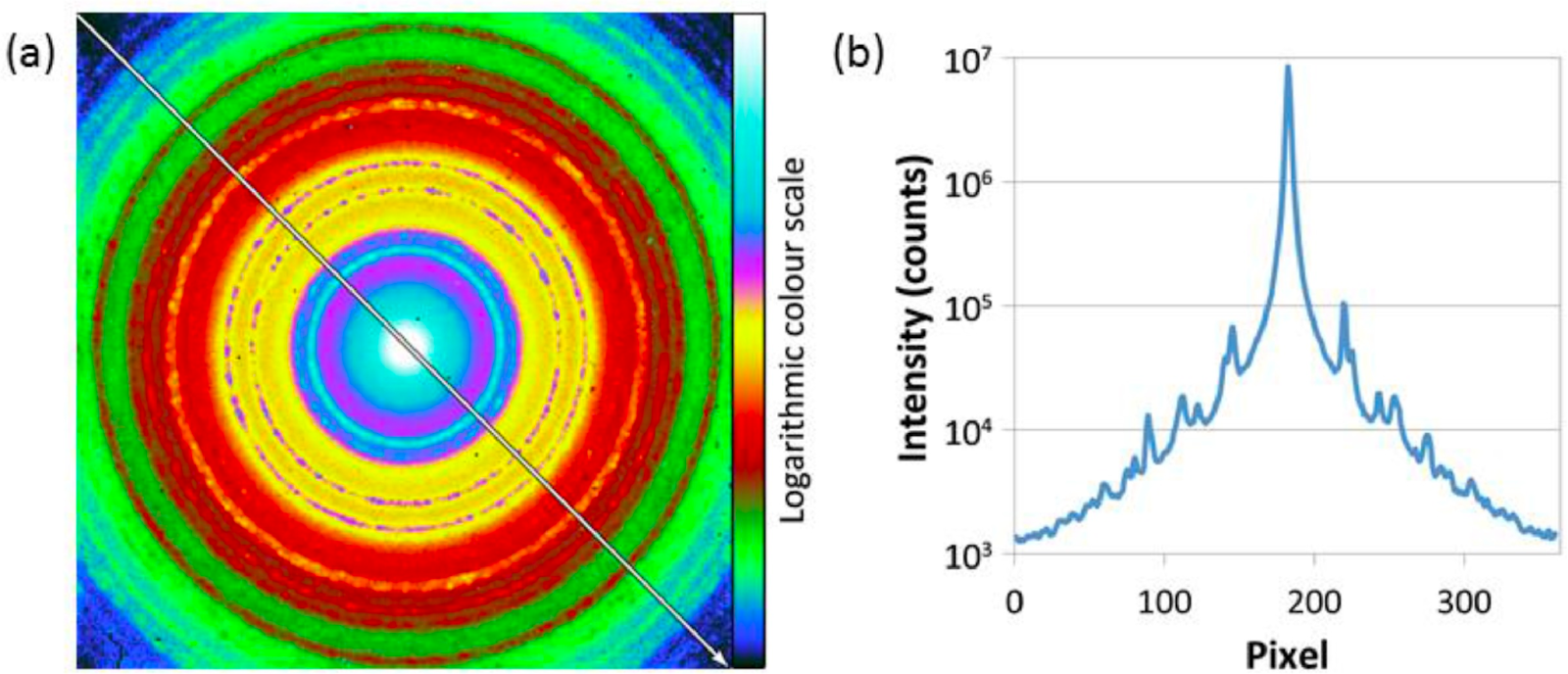
In diffraction, MerlinEM can be used to image direct probes while still retaining the ability to count single electrons for the weaker diffraction spots. This makes the detector an attractive solution for low-dose techniques in the microscopy of bio-molecules, microED and scanning precession techniques.
In EELS, the 4x1 MerlinEELS detector version, offers low noise, large dynamic range and fast framing. These characteristics are can be used to push core-loss imaging to high kV losses [5] and allow live drift corrections.
- References
References:
[1] Mac Raighne, A. et al., Journal of Instrumentation 6.01 (2011): C01047.
[2] Mir, J. A. et al., Ultramicroscopy 182 (2017): 44-53.
[3] Krajnak, M. et al., Ultramicroscopy 165 (2016): 42-50.
[4] Ning, S. et al., Microscopy and Microanalysis (2022): 1-11.
[5] Tencé, M. et al., Microscopy and Microanalysis, 26(S2), 1940-1942.