Detection under €1000 - Creating a sustainable, cost-effective electron detector
- Abstract number
- 261
- Presentation Form
- Poster
- Corresponding Email
- [email protected]
- Session
- Poster Session One
- Authors
- Mr Cameron O'Byrne (1), Mr Sean Fulham (1), Dr Jonathan Peters (1), Dr Lewys Jones (1)
- Affiliations
-
1. Trinity College Dublin
- Keywords
SiPM, YAG scintillators, Symmetrical charge division circuit, 4x4 array, low cost, sustainable
- Abstract text
For early-career researchers or scientists in smaller labs, the cost of equipment is a significant barrier to entry into the world of electron microscopy. Reducing this barrier would bring wider access to the latest microscopy techniques and greater democratisation of science, furthering the investigation of research projects from a vast array of fields.
Here, we set out to create a low-cost and easily reproducible electron detector for a scanning transmission electron microscope (STEM) using a repurposed SiPM array component and an inorganic scintillator. SiPMs are highly sensitive and can detect light in a wide range of wavelengths, making them ideally suited to scintillator-based photon detection. A Broadcom AFBR 4x4 pixel SiPM was used for the detector due to its fast rise time in detection.
An asymmetrical charge division circuit 1, was designed to reduce the signal outputs of the detector from 16 to 8 while also including an amplification element to increase the signal output from the SiPM. The PCB board, fig 1, was designed to be two-sided tile-able to allow for the possibility of increasing the 4x4 array to an 8x8 by adding three identical detectors together in a square pattern.

Fig 1 – The two sides of the board of the 4x4 array detector. The SiPM was placed in the corner of the PCB to enable it to be two side tile-able. The electrical components were kept on the back side of the PCB to reduce electron beam interference.
A thin layer of YAG scintillator crystal was placed on top of the SiPM array to convert the energy of the incoming electrons into photons. This was acquired in a larger sheet and cut to exactly fit the surface area of the SiPM using a laser engraver.
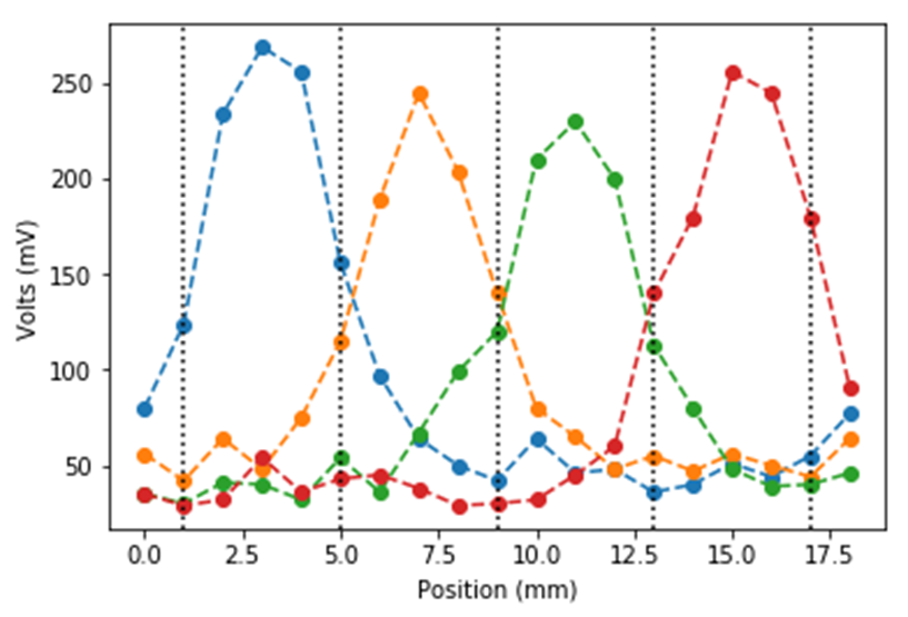
Initial tests were performed in the detector, Fig 2, with just a single row of the array being used linearly. Each Section of the four-part row was then individually read out as the detector was translated across horizontally, giving the sensitivity of each pixel in the row.
This detector is intended to cost a fraction of the price of the current detectors, and although perhaps limited in some aspects, it provide for a gap in the current electron detection market. They representthe ground floor of the future development of more detectors with ever-increasing quality.
Fig 2 – The Readout from four linear sections of the SiPM detector. Each colour represents a pixel readout and the dashed lines are the physical boundaries between them
Acknowledgments:
The authors would like to acknowledge the Advanced Microscopy Lab for assisting in this work and providing infrastructural support. C. O'Byrne acknowledges AMBER grant 17/RC-PhD/3477, J. Peters acknowledges SFI grant 19/FFP/6813, and L. Jones acknowledges SFI URF grant URF/RI/191637.
- References
1 - Stratos D, Maria G, Eleftherios F et al. - Comparison of three resistor network division circuits for the readout of 4x4 pixel SiPM arrays. (2013). DOI: 10.1016/J.NIMA.2012.08.006