Aberration-Corrected Integrated Center of Mass (iCOM) Imaging
- Abstract number
- 44
- Presentation Form
- Poster
- DOI
- 10.22443/rms.mmc2023.44
- Corresponding Email
- [email protected]
- Session
- EMAG - 3D & Tomographic Electron Microscopy
- Authors
- Mr. Zhiyuan Ding (1), Prof. Angus Kirkland (1, 2, 3), Prof. Peter Nellist (1)
- Affiliations
-
1. Department of Materials, University of Oxford
2. Electron Physical Sciences Imaging Centre, Diamond Light Source Ltd., Harwell Science and Innovation Campus
3. The Rosalind Franklin Institute, Harwell Campus
- Keywords
transmission electron microscopy, iCOM, ptychography, single-side band, aberration correction
- Abstract text
Introduction
Integrated center of mass (iCOM) is a commonly used phase-contrast imaging method in scanning transmission electron microscopes (STEM). The contrast in iCOM comes from the movement of center of mass (COM) of a convergent beam electron diffraction pattern (CBED). This movement is related to the deflection of electron beam by the static electric field of a sample. It has been shown that the contrast of the iCOM image is proportional to the phase change of the exit wave resulting from the projected potential of the sample, which makes iCOM a phase-contrast imaging method, if a phase object approximation (POA) is assumed [1].
Here we report a new method, which can correct residual aberrations in iCOM images, named Single-Side Band masked iCOM (SSBm-iCOM) to calculate a filtered iCOM image from a 4DSTEM dataset. In this method, the Fourier transform of COM mapping is calculated in reciprocal space and a mask which is similar to the mask used in Single-Side Band (SSB) ptychography [2, 3] is applied. The reason we apply a SSB mask is that, theoretically, if the sample can be regarded as a weak phase object, then all phase-contrast information is included in the SSB mask [4]. Therefore, this mask optimally filters the original dataset and removes pixels without phase-contrast information and that just contain noise, and compensate the residual aberration in 4DSTEM dataset.
Simulation
We simulated SSBm-iCOM and conventional iCOM using the same imaging conditions and sample (A monolayer graphene sample with an atom replaced by silicon). The 4DSTEM dataset is generated using a modified script based on MULTEM [5].
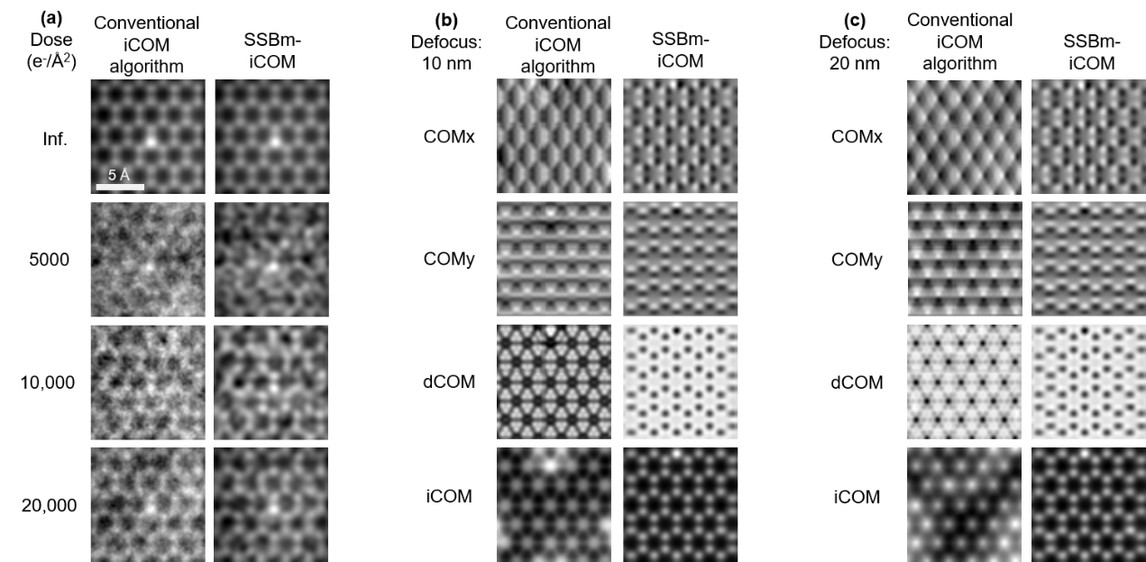
Simulations were carried out at 80 kV acceleration voltage with a convergence angle of 22.5 mrad (Figure 1a), 30 mrad (Figure 1b-c) and a scan step of 0.2 Å. The overall collection angle of detector was 128 mrad with a detector size of 256×256 pixels. Poisson noise was applied to simulate low-dose conditions from 5×10³ to 2×10⁴ e-/Å2 (Figure 1a), where SSBm-iCOM images show less noisy phase-contrast images, and better lattice structures. This is because the SSB mask filters unnecessary signals which actually “increase” the signal-to-noise ratio of low-dose 4DSTEM datasets. Reconstructions of defocus datasets (10 nm for Figure 1b and 20 nm for Figure 1c) show the residual aberrations are compensated.
Figure 1 | Simulations of conventional iCOM algorithm and SSBm-iCOM for (a) different dose conditions, (b) aberration correction with a defocus of 10 nm, (c) aberration correction with a defocus of 20 nm.
Conclusions and Future work
Single-side band masked iCOM (SSBm-iCOM) is a phase-contrast imaging method that combines the features of SSB ptychography and traditional iCOM, but can correct residual aberrations in 4DSTEM datasets and has good tolerance to low-dose conditions.
- References
[1] I. Lazić, E. G. Bosch, and S. Lazar, "Phase contrast STEM for thin samples: Integrated differential phase contrast," Ultramicroscopy, vol. 160, pp. 265-280, 2016.
[2] C. M. O’Leary, G. T. Martinez, E. Liberti, M. J. Humphry, A. I. Kirkland, and P. D. Nellist, "Contrast transfer and noise considerations in focused-probe electron ptychography," Ultramicroscopy, vol. 221, p. 113189, 2021.
[3] J. Rodenburg, B. McCallum, and P. Nellist, "Experimental tests on double-resolution coherent imaging via STEM," Ultramicroscopy, vol. 48, no. 3, pp. 304-314, 1993.
[4] T. J. Pennycook, A. R. Lupini, H. Yang, M. F. Murfitt, L. Jones, and P. D. Nellist, "Efficient phase contrast imaging in STEM using a pixelated detector. Part 1: Experimental demonstration at atomic resolution," Ultramicroscopy, vol. 151, pp. 160-167, 2015.
[5] I. Lobato and D. Van Dyck, "MULTEM: A new multislice program to perform accurate and fast electron diffraction and imaging simulations using Graphics Processing Units with CUDA," Ultramicroscopy, vol. 156, pp. 9-17, 2015.