Correlation of nanoscale electromechanical and mechanical properties of twisted double bi-layer graphene via UFM, PFM, and E-HFM
- Abstract number
- 403
- Presentation Form
- Poster
- DOI
- 10.22443/rms.mmc2023.403
- Corresponding Email
- [email protected]
- Session
- Poster Session Two
- Authors
- Sergio Gonzalez-Munoz (1), Alessandra Canetta (2), Dr. Khushboo Agarwal (1), Dr. Jean Spièce (2), Prof. Pascal Gehring (2), Prof. Oleg V. Kolosov (1)
- Affiliations
-
1. Lancaster University
2. Université Catholique de Louvain
- Keywords
Electromechanical, twistronics, graphene, Young's modulus
- Abstract text
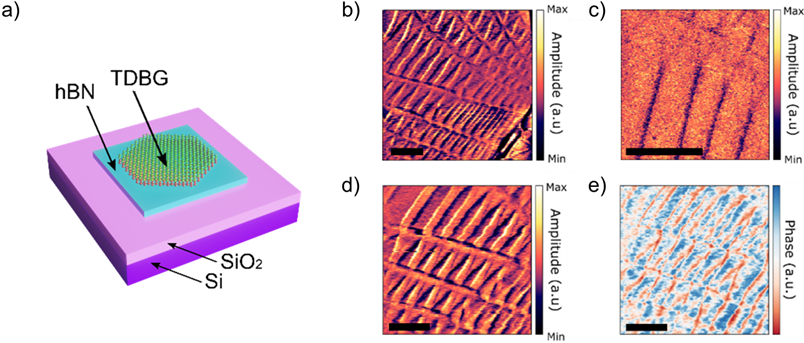
Recently, multiple theoretical and experimental studies have been published regarding the properties of stacked two-dimensional (2D) layers forming a twisted heterostructure. This field (known as twistronics) shows that properties of 2D materials can be modified to a great degree, including bandgap modulation and creating superconductive structures. Given the versatility that these structures have, many exciting engineering is being applied to them resulting in promising properties.
In this study, we investigated a heterostructure composed of two twisted graphene bi-layers with a small angle between them (1.1º), where an atomic reconstruction is induced changing the lattice symmetry and creating a Moiré pattern. The electrical and mechanical properties of the 2D nanostructure are affected by this symmetry reconstruction, generating relaxation-induced strain gradients. We compared nanomechanical mapping via Ultrasonic Force Microscopy (UFM) and electromechanical response probed by Piezoresponse Force Microscopy (PFM) and Electrical Heterodyne Force Microscopy (E-HFM). These allowed us to assign Moiré patterns of the heterostructure to the particular crystallographic arrangements and to quantify the local Young’s modulus variation between single and double domain walls. Moreover, by measuring these domain walls specifically with PFM, it is possible to extract evidence of non-uniform strain in stretched triangular domains in the Moiré pattern. The phase images from the E-HFM allow us to observe a fast time-domain nanoelectromechanical relaxation in the order of picoseconds with nanoscale lateral resolution.
Figure 1. a) Structure of the measured sample. b) Piezoelectric Force Microscopy, c) Ultrasound Force Microscopy, and e), f) Electric-Heterodyne Force Microscopy amplitude and phase, respectively. Scalebars: 200 nm.