Passive identification of adsorbate single bomobenzene molecules on atomically resolved Si(111)-7×7 surface by I-z spectroscopy at room temperature
- Abstract number
- 90
- Presentation Form
- Poster & Flash Talk
- DOI
- 10.22443/rms.mmc2023.90
- Corresponding Email
- [email protected]
- Session
- Atomic and Molecular Resolution Phenomena via AFM, STM and Scanning Probes
- Authors
- Mr Pieter Keenan (2, 1, 3), Mr Thomas Coates-Evans (3), Mr Shean Ong (3), Dr Peter Sloan (1, 3), Dr Kristina Rusimova (2, 1, 3)
- Affiliations
-
1. Centre for Nanoscience and Nanotechnology, University of Bath
2. Centre for Photonics and Photonic Materials, University of Bath
3. Department of Physics, University of Bath
- Keywords
STM, Single molecule, Manipulation, Desorption, Surfaces
- Abstract text
Summary: The identification of the nature of dark spots in an STM image is important to single molecule manipulation probability determination. This study demonstrates how adatom vacancies on the Si(111)-7x7 surface can be distinguished from possible desorption sites in a room-temperature STM via passive I-z spectroscopy measurements of the tunnelling current decay constant. This will improve analysis of STM manipulation experiments by removing dark spots from which manipulation is not possible from the dataset.
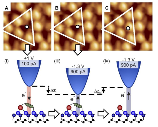
A surface’s local density of states (LDoS) can be probed with atomic resolution using scanning tunnelling microscopy. This produces a topographical map of ‘bright’ and ‘dark’ spots corresponding to the position of the atoms on the surface. On the metallic Si(111)-7x7 surface, the Si adatoms appear as bright spots due to their high LDoS. Dark spots correspond to either adatom vacancies, contaminants, or molecules intentionally chemisorbed onto the surface (Figure 1a).
Figure 1: High-resolution STM images and (below) corresponding schematic diagrams of a manipulation procedure (imaging parameters +1 V, 100 pA, 3 nm by 3 nm). (A) Before manipulation image: a half unit cell of Si(111)-7×7 is outlined, the white circle atop the missing-adatom like dark spot location indicates the position of a single aromatic molecule. (B) During image, (C) after manipulation image showing a bright adatom revealed in place of the dark spot.
Single molecule manipulation experiments inducing the desorption of either single or multiple aromatic molecules via charge injection into a Si(111)-7x7 surface from the tip of a scanning tunnelling microscope (STM) are well established [1- 4]. In such experiments, STM images taken before and after current injection with passive imaging parameters are compared. Locations from which molecules have been successfully desorbed reveal a bright Si adatom where a dark spot had previously been (Figure 1). Unsuccessful desorption experiments are then taken to be those where a dark spot remains in the ‘after’ STM image.
Given the relatively small number of measurements made in a typical STM-induced manipulation dataset (O(10-100)), incorrectly identifying a vacancy or contaminant dark spot as a molecule can significantly impact reported manipulation probabilities. Therefore, this study aimed to develop a passive technique to differentiate molecules from other dark spots with a single measurement in a UHV, room temperature STM.A simple 1D model of an STM gives the tunnelling current (I) as a function of tip-sample separation (z) as
I = Ae−2κz ,
where A is a constant of proportionality and the decay constant κ is dependent on the properties of the sample beneath the STM tip. An expression for κ can be found by taking the derivative of the current, such that
κ = − 1/2I(∂I/∂z).
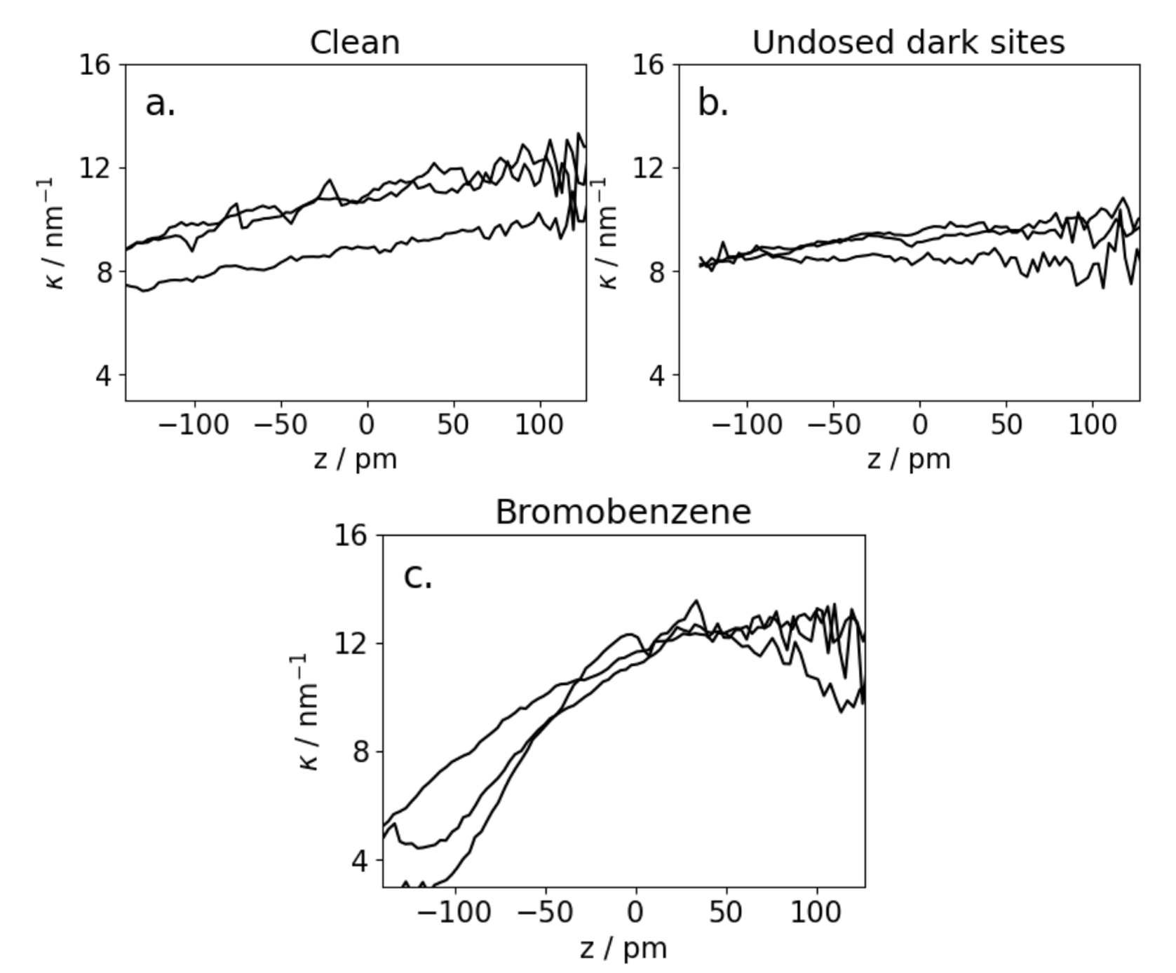
With a lock-in amplifier to approximate ∂I/∂z , sweeps of κ(z) have been measured for faulted corner adatom locations in the Si(111)-7x7 unit cell. Experiments were performed above clean adatom sites, dark spots on a undosed surface, and dark spots on a surface dosed with bromobenzene.
After measurement of κ above dark spots on the dosed surface, 750 pA of current was injected into the surface for 120 s at a bias voltage of -2.1 V. These are parameters known to cause non-local manipulation of bromobenzene [3], leading to desporption of molecules within a radius of the tip location. Comparison of before and after passive scan images allowed identification of the κ(z) measurements that correspond to bromobenzene desorption locations.
Values of κ(z) relative to a set-point height (defined as 0) corresponding to a tunnelling current of 100 pA at a bias of +1 V are displayed in Figure 2. Every clean site measurement displayed a gently sloping featureless κ(z) curve, with the undosed dark spots also showing a similar trend. There was no desorption from any sites with the featureless κ(z) curve over the swept z region. All the sites at which a desorption event was identified showed a marked decrease in κ at small z.
Figure 2: Representative κ(z) curves recorded at faulted corner adatom sites for a) clean adatoms, b) dark spots on an undosed surface and c) dark spots on a bromobenzene dosed surface that became bright after a desorption experiment. Each curve was recorded with a bias of +1 V, a settling time of 10 ms and lock-in averaging time of 100 ms at each of the 160 data points.
It was therefore concluded that any dark faulted corner site at which a flat κ(z) curve is observed does not correspond to an adsorbed bromobenzene molecule. Noting the κ(z) plots are similar for the clean and undosed dark cases, that Si adatom vacancies are common on the surface [6] and the UHV conditions, it is suggested that the dark spots with flat κ(z) profiles are Si adatom vacancies. In the vacancy hypothesis, the measured κ(z) curve would be a convolution of the LDoS of the Si atom located a layer down with contributions from the adatoms surrounding the vacancy, which would result in the clean adatom-like profile observed. More importantly, this experiment has shown that dark spots from which desorption is possible can be distinguished from other dark spots as having qualitatively different κ(z) curves in a passive STM measurement. We believe such dark spots with this κ(z) profile are likely bromobenzene, but further work with other contaminant molecules is required to verify this. The technique developed here will allow a subset of dark spots that might previously have been identified as candidates for a desorption experiment to be removed from consideration in the analysis.
Passive I-z spectroscopy measurements of the tunnelling current decay constant κ show qualitative differences for STM image dark spots at sites from which molecular desorption reactions are observed. This technique differentiates between what are believed to be Si adatom vacancies and sites from which molecular desorption is possible. This will improve the analysis of STM manipulation experiments by removing dark spots from which manipulation is not possible from the dataset.
- References
1] Kristina R Rusimova and Peter A Sloan. “Molecular and atomic manipulation mediated by electronic excitation of the underlying Si(111)-7x7 surface”. In: Nanotechnology 28.5 (Dec. 2016), p. 054002. doi: 10.1088/1361-6528/28/5/054002. url: https://dx.doi.org/10.1088/1361-6528/28/5/054002.
[2] K. R. Rusimova et al. “Initiating and imaging the coherent surface dynamics of charge carriers in real space”. In: Nature Communications 7.1 (Sept. 2016), p. 12839. issn: 2041-1723. doi:10.1038/ncomms12839. url: https://doi.org/10.1038/ncomms12839.
[3] H G Etheridge, K R Rusimova, and P A Sloan. “The nanometre limits of ballistic and diffusive hot-hole mediated nonlocal molecular manipulation”. In: Nanotechnology 31.10 (Dec. 2019), p. 105401. doi: 10.1088/1361-6528/ab5d7c. url: https://dx.doi.org/10.1088/1361-6528/ab5d7c.
[4] Peter A Sloan and Kristina R Rusimova. “A self-consistent model to link surface electronic band structure to the voltage dependence of hot electron induced molecular nanoprobe experiments”. In: Nanoscale Advances 4.22 (2022), pp. 4880–4885.
[6] Zsolt Majzik et al. “Room Temperature Discrimination of Adsorbed Molecules and Attachment Sites on the Si(111)–7 × 7 Surface Using a qPlus Sensor”. In: ACS Nano 7.3 (Mar. 2013), pp. 2686–2692. issn: 1936-0851. doi: 10.1021/nn400102m. url: https://doi.org/10.1021/nn400102m.