Remote-refocus microscopy using a MEMS piston micromirror
- Abstract number
- 199
- Presentation Form
- Poster
- DOI
- 10.22443/rms.mmc2023.199
- Corresponding Email
- [email protected]
- Session
- Poster Session Two
- Authors
- Mr Jay Christopher (1), Dr Liam Rooney (2), Professor Deepak Uttamchandani (1), Dr Ralf Bauer (1)
- Affiliations
-
1. Department of Electronic and Electrical Engineering, University of Strathclyde
2. Strathclyde Institute of Pharmacy & Biomedical Sciences, University of Strathclyde
- Keywords
Remote-refocusing, microelectromechanical systems, MEMS
- Abstract text
Summary
We demonstrate a remote-refocus microscopy system with volume acquisition rates of up to 150 Hz using a microelectromechanical systems (MEMS) mirror with controllable piston movement. The small footprint, affordable and easily controllable 2 mm mirror allows nm scale alignment precision of the focal plane, with an overall axial focus shift of up to 225 µm with a 100x 1.25 NA imaging objective.
Introduction
Volumetric imaging and focal adjustment is in many systems done using stage scanning or piezo objective scanners with the speed in these cases limited by the mass of a stage/objective and by the potential of sample disturbances in media coupled objectives. The use of electrical tunable lenses or the remote-refocus approach first demonstrated by Bouchard et al.[1] can circumvent this limitation, with specifically remote-refocus approaches allowing a fixed objective-sample system without deterioration of the image quality. Their basic approach re-images the main microscope path through a secondary and potentially tertiary microscope, with sample focal shifts achieved by moving a reflective mirror in a 2-objective configuration or the 3rd objective in a 3-objective configuration[2]. Iterations using piezoelectric stages, linear translation stages or adaptive optics mirrors[3] have so far been shown. We will demonstrate a setup with a simple and accurate control through a Microelectromechanical Systems (MEMS) device with tip/tilt and piston movement potential.
Materials and Methods
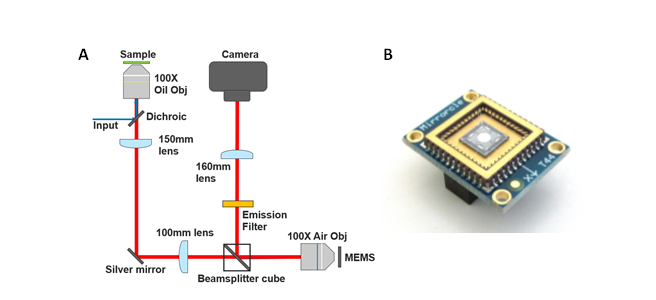
The remote-refocus microscope setup is built around a combination of a Zeiss 100x 1.25NA oil and 0.9NA air objective in the sample and remote space, respectively, with a reflection configuration in the remote space enabling the use of a reflective MEMS mirror for fast focal control. A schematic of the setup is shown in Figure 1(a).
The objectives are correlated with tube lenses of 150 mm and 100 mm focal length to satisfy the remote-refocus condition for aberration free remote imaging in fixed samples with refractive index around 1.5. Brightfield white-light illumination and widefield epifluorescence illumination are used to demonstrate the volume imaging speed and characteristics. A 160 mm final tube lens and sCMOS camera (Prime BSI Express) are used for image collection.
Figure 1 - (A) Schematic of remote-refocus imaging system and (B) Mirrorcle MEMS example.
The 3-axis MEMS mirror (see Figure 1(b)) is characterised using a single-point vibrometer with nanometer piston measurement accuracy to evaluate the mirror step response behaviour as well as the response to dynamic sinusoidal or ramp position changes.
Results and Discussion
The MEMS piston motion shows a step response of <4 ms for position changes ranging from 10 nm to the maximum piston motion of 75 µm. Sinusoidal input position control voltages are followed accurately, with a 20 % movement amplitude drop-off at 200 Hz, originating from the use of a 500 Hz electronic filter to reduce unwanted mirror oscillations. Ramp position control voltages with fast fly-back with <4 ms fly-back time are accurately reproduced up to a ramp frequency of 150 Hz, after which movement oscillations are present at the mirror turning points.
Brightfield images of a USAF resolution test target are used to confirm telecentric imaging and calibrate camera pixel sizes. Remote-refocusing and imaging performance is evaluated using 175 nm fluorescence beads mounted in a volume of ProLong Glass (Invitrogen P36982), showing a volumetric imaging possibility with 300 nm resolution over the full addressable z-range of the MEMS. 3D live-cell sample images of fluorescent test specimens will be shown, evaluating the potential of >100 Hz full volumetric imaging with the MEMS remote-refocus setup as well as a fast, arbitrary focal plane selection mode.
Conclusions
We show the implementation and characterisation of a remote-refocus microscopy setup making use of an affordable MEMS micromirror with piston and tip/tilt movement potential to enhance a remote-refocus volume imaging speed potential due to the low mass of the MEMS used as the focal control element.
- References
References
[1] E. J. Botcherby, R. Juškaitis, M. J. Booth, and T. Wilson, “An optical technique for remote focusing in microscopy,” Opt. Commun., vol. 281, no. 4, pp. 880–887, 2008, doi: 10.1016/j.optcom.2007.10.007
[2] M. Gintoli et al., “Spinning disk-remote focusing microscopy,” Biomed. Opt. Express, vol. 11, no. 6, p. 2874, 2020, doi: 10.1364/boe.389904.
[3] T. Wright, H. Sparks, C. Paterson, and C. Dunsby, “Video-rate remote refocusing through continuous oscillation of a membrane deformable mirror,” JPhys Photonics, vol. 3, no. 4, 2021, doi: 10.1088/2515-7647/ac29a2.
Acknowledgements
We acknowledge funding from the UK Engineering and Physical Sciences Research Council (grant EP/S032606/1, studentship EP/T517938/1), The Leverhulme Trust, and UK Royal Academy of Engineering (Engineering for Development Fellowship scheme RF1516/15/8).