Microscopic analysis of new and historic cotton fibre cross-sections
- Abstract number
- 369
- Presentation Form
- Poster & Flash Talk
- Corresponding Email
- [email protected]
- Session
- Analysis and Imaging in Heritage Science
- Authors
- Miss Rana T A Salem (1), Mrs Karen Thompson (2), Dr Mahesh Uttamlal (1)
- Affiliations
-
1. Glasgow Caledonian University
2. University of Glasgow
- Keywords
AFM, SEM, wide field optical microscopy, cotton, fibres, nano-structure, heritage science, textile conservation.
- Abstract text
The presentation describe the microscopic analysis of new and historic cotton fibres cross- pre- and post-bleaching; methods commonly employed by museums for conservation of historic textile materials.
All historical textile materials (including tapestries, carpets, decorated textiles, and clothing), most commonly protein and/or cellulose in composition, will degrade over time due to exposure to dirt, microorganisms and environmental conditions, i.e., humidity, oxygen and pollutants [1]. Conservators in museums throughout the world are responsible for the care of these textiles, which are important sources of cultural heritage.
The conservation process is generally determined by the condition of the textile fibre, dye composition, and age, construction, as well as the object’s history of use and storage conditions. For cellulose textiles, aqueous immersion techniques can be used for removing stains and yellowing using both oxidizing (e.g., H2O2) and reducing (e.g., NaBH4) agents.
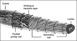
The aim of this project is to assess the assess the nanostructure of cellulose (cotton) fibres (Figure 1) from new and historic textiles by examining the cross-sections pre-and post-cleaning using scanning electron microscopy and atomic force microscopy. This work is an extension to our previous work looking at the impact of bleaching on the outer surface of the fibres [2][3].
Figure 1: Schematic diagram of the cotton fibre structure.
Raw calico, scoured (Whaleys Ltd) and historic (shirt circa. 1900) cotton textiles were tested. Cross sections of cotton threads taken from the samples were prepared by setting in resin then slicing using a microtome directly onto microscope slides and SEM stubs. The samples were all 1µm thick.
Analysis of the cross sections were performed using optical microscopy (Leica DMR, Leica Microsystems Ltd), scanning electron microscopy (EVO 50 XVP, Carl Zeiss SMT Ltd) and atomic force microscopy (Dimension 3100 (Veeco Metrology) Nanoscope IV Scanning Probe Microscope).
The data presented in this work were obtained from samples pre-bleaching. No post-bleaching data will be presented here.
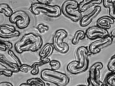
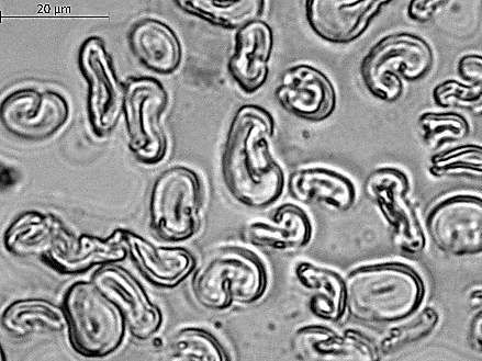
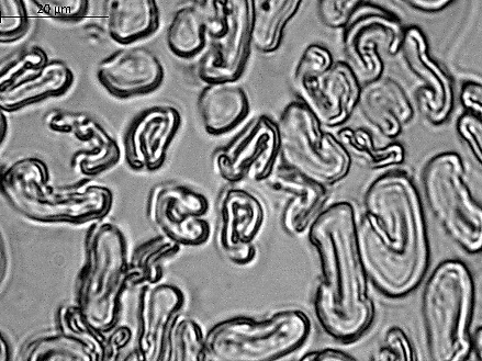
Brightfield images of cross-sections of raw, scoured and historic samples are shown in Figure 2. Evidence of disruption to the integrity of the fibres is noted for the historic fibres, however, the resolving power limits the overall assessment of the fibre condition. It is difficult to distinguish between the samples.
Figure 2: Optical microscopy of cotton fibre cross sections - (left) raw, (middle) scoured and (right) historic. Images were taken using a ×100 plan achromat objective.
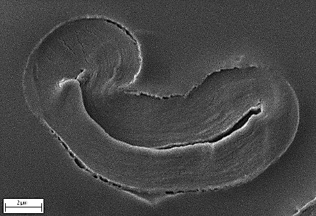
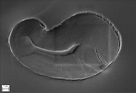
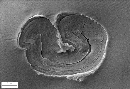
SEM (SE1) micrographs of cotton fibre cross-sections are shown in Figure 3. Typically, we find intact wall structure in the raw cotton samples however, the presence of cracks in the scoured samples are seen. Historic samples typically show more cracking and disruption to the secondary wall.
Figure 3: SEM (SE1) micrographs of cross sections of cotton fibres - (left) raw, (middle) scoured and (right) historic. (EHT = 5kV, Samples Au/Pd coated.)
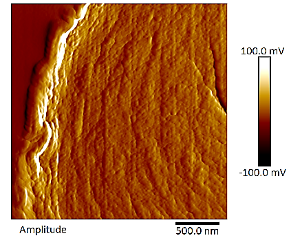
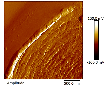
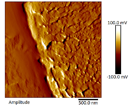
Typical AFM amplitude images of fibre cross-sections are shown in Figure 4. Images show the outer wall layers and the secondary wall. The raw sample show a fully intact outer cuticle and primary walls whereas the scoured samples shows a thinner wall as a result of scouring. The historic sample shows large amounts of disruption to the outer and inner walls.
Figure 4: AFM amplitude images of cotton fibre cross-section showing outer wall - (left) raw, (middle) scoured and (right) historic sample. (Image size - 2.5 × 2.5 µm)
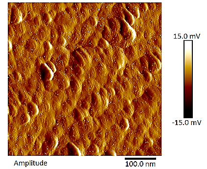
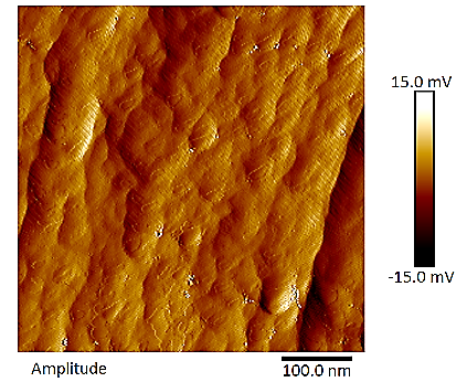
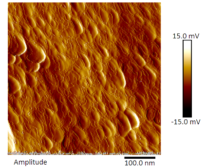
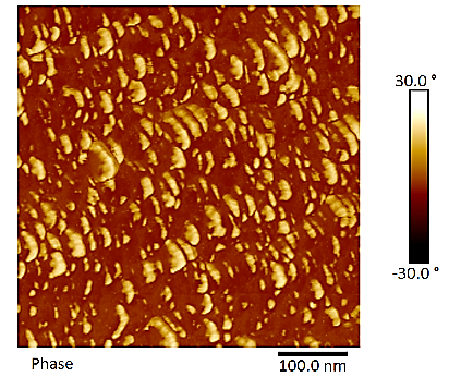
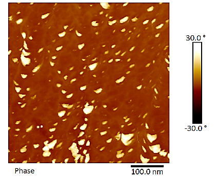
Figure 5 shows typical amplitude and phases images of the secondary wall of all three samples. It is possible to see the nano-fibrils that make up the secondary wall. The amplitude images show the compactness of the fibrils. The fibrils are generally circular to oblong in shape. The phase images give an indication of the elastic properties of nano-fibrils. The historic sample is shown to be much softer compared to the raw and scoured samples.
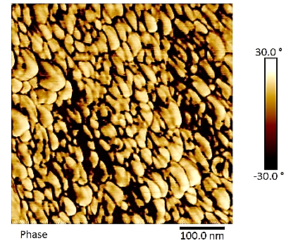
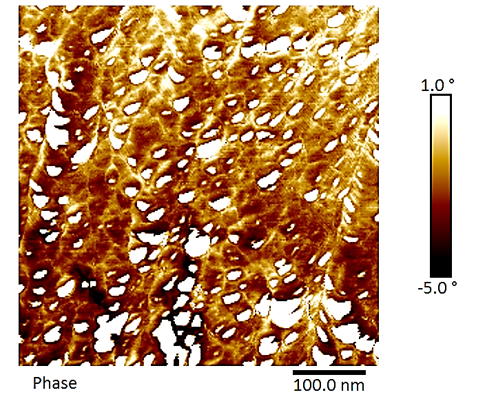
A contrast enhanced phase image of the scoured cotton cross-section is shown in Figure 6. This image shows, for each fibril, there is an outer and inner component. The two components vary in their elastic properties and may indicate the presence of both crystalline and amorphous cellulose.
Figure 5: AFM (top) amplitude and (bottom) phase images of cross section of cotton fibre inner secondary wall, (left) raw, (middle) scoured and (right) historic sample. (Image size - 500 × 500 nm)
Figure 6: Contrast enhanced AFM phase image of scoured cotton showing bilayer structure of the nano-fibrils.
In conclusion, the use of multiple imaging techniques has shown that it is possible to analyse the micro and nano-structure of cotton fibres. The information can be used to assess the integrity and quality of the fibres. This information can be of importance and have implications for textile conservation and sustainability throughout the world.
- References
[1] Bresee RR. General effects of ageing on textiles. J Am Inst Conserv. 25(1) 39 (1986) . https://doi.org/10.1179/019713686806028032.
[2] Rana T. A. Salem, Mahesh Uttamlal and Karen Thompson, Microscopic analysis of new and historic cotton textiles pre- and post-bleaching, Microscopy Microscience Congress 2021. Available on-line: https://www.mmc-series.org.uk/mmc2021/abstract/microscopic-analysis-of-new-and-historic-cottontextiles-pre-and-post-bleaching.html
[3] Salem, R.T.A., Thompson, K. & Uttamlal, M. Bleaching cotton in textile conservation: a closer look using atomic force microscopy. Herit Sci 10, 195 (2022). https://doi.org/10.1186/s40494-022-00830-2