Developing 3D standards for super-resolution imaging.
- Abstract number
- 552
- Presentation Form
- Poster
- DOI
- 10.22443/rms.mmc2023.552
- Corresponding Email
- [email protected]
- Session
- Poster Session Three
- Authors
- Ms Tina Sarapa (1), Dr Colin Rickman (1)
- Affiliations
-
1. Heriot-Watt university
- Keywords
SMLM, Super-resolution imaging, 3D microscopy, PSF, beads, astigmatism, biplane imaging.
- Abstract text
Super-resolution imaging refers to a well-established set of microscopy techniques that allow researchers to bypass the limit of resolution of optical microscopy. Single molecule localisation microscopy, or SMLM, is a set of techniques that are based on separating individual point spread functions (PSF) in space. The structures of interest within a sample are labelled with fluorophores that blink (either spontaneously or controlled depending on the specific technique). The sample is usually imaged over time, and the resulting frames are used to digitally reconstruct a super-resolved image. Most commonly SMLM is performed in 2D, however several 3D techniques have been developed recently, including the use of astigmatic lenses, biplane imaging, PSF double helix, and other methods. Most are based on manipulating PSFs to allow determination of molecular position in the Z-axis. All of these 3D techniques require calibration standards, which our research group has been working on. Specifically, we have focused on astigmatism and biplane imaging techniques.
Two general types of errors typically occur during 3D imaging. The first type is the localisation precision error, which depends on the signal-to-noise ratio and determines the accuracy of localising a single molecule. The second type is how accurate the localised molecular positions describe to the underlying structure. Localisation precision is empirically determined during the localisation process based on photon statistics. However, to determine the ability of SMLM to accurately reproduce a structure, standards that establish a ground truth for error calculation are required. We have developed a 3D structure of known dimensions using a chemically reactive bead with an impermeable surface decorated with fluorophores. This results in a thin, defined curved surface in three dimensions with a known shape over a large range of axial position. A range of fluorophores can be conjugated to the beads allowing the fluorophore and imaging parameters to be matched to the experimental imaging conditions. Because of this, such standards will be widely applicable to a range of super-resolution techniques, such as STED, PALM, D-STORM and others.
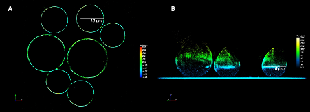
Figure 1: Panel A shows the equatorial cross-section of beads labelled with Alexa 647 in MEA. Panel B shows the reconstructed Z profile of the same beads, coloured by the pixel depth. The data was obtained on the Bruker Vutara VXL biplane imaging system.
- References
Winterflood CM, Platonova E, Albrecht D, Ewers H. Dual-color 3D superresolution microscopy by combined spectral-demixing and biplane imaging. Biophys J. 2015 Jul 7;109(1):3-6. doi: 10.1016/j.bpj.2015.05.026. PMID: 26153696; PMCID: PMC4571034.
Cabriel, C., Bourg, N., Jouchet, P. et al. Combining 3D single molecule localization strategies for reproducible bioimaging. Nat Commun 10, 1980 (2019). https://doi.org/10.1038/s41467-019-09901-8