Fluorescence microscopy for everyone: epifluorescence imaging with the OpenFlexure Delta Stage
- Abstract number
- 559
- Presentation Form
- Poster
- DOI
- 10.22443/rms.mmc2023.559
- Corresponding Email
- [email protected]
- Session
- Poster Session Three
- Authors
- Freya Whiteford (2), Dr Samuel McDermott (1), Dr Joe Knapper (2), Dr Richard Bowman (2)
- Affiliations
-
1. University of Cambridge
2. University of Glasgow
- Keywords
OpenFlexure, Delta Stage, accessible microscopy, open source, global health, fluorescence, epifluorescence, 3D printing, superresolution, structured illumination
- Abstract text
High quality microscopes and positioning stages are vital to biological and medical research but can require significant financial investment to acquire and maintain in working condition, leading to inequality in accessibility. The OpenFlexure family of open-source microscopes and positioning stages aim to alleviate this via 3D-printed, flexure-based low cost designs that can be assembled and maintained by their users.
Customisation is a particular strength of the OpenFlexure designs, as the open-source nature of the project allows hardware to be adapted to specific imaging modalities and use cases as needed. For instance, support for RMS objectives is provided alongside modifications for less expensive imaging optics.
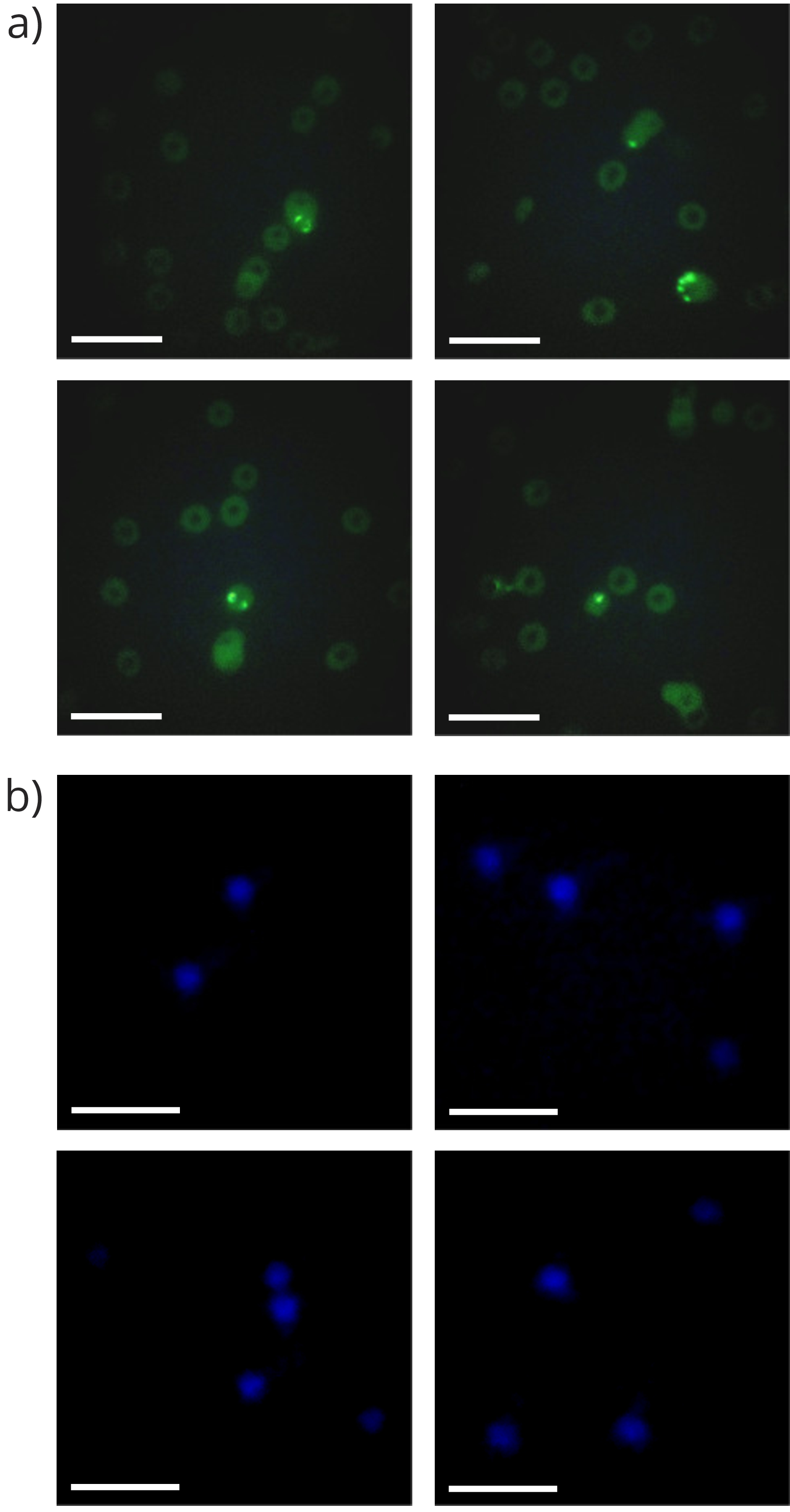
Fig 1. The OpenFlexure Delta Stage is capable of imaging samples with quite low fluorescent intensity. (a) Epi-fluorescence micrographs of SYBR-green stained malaria infected red blood cells show the location of the red blood cells and the brighter regions show the locations of the malaria parasites. It is possible to see the characteristic ring shape of the trophozoite stage of the malaria erythrocytic cycle. (b) Epi-fluorescence micrographs show 6 µm fluorescent dyed ~6 µm microspheres. They are illuminated with the UV LED and emit blue light. Images cropped and contrast enhanced for visibility, scale bars 20 µm. Reproduced from [1].
Fluorescence microscopes are especially useful for providing contrast when imaging biological samples. The OpenFlexure Delta Stage [1] is a 3D-printed flexure stage with a non-Cartesian actuator alignment, allowing the stage to move independently of the microscope optics in 3 dimensions. Following on from an earlier design for an epi-fluorescence reflection microscope using the OpenFlexure Delta Stage [1], we present refinements to the Delta Stage fluorescence microscope for further ease of assembly as well as a wider range of filter cubes and LEDs.
As well as epifluorescence, the OpenFlexure Delta Stage is also capable of trans-illumination and polarisation contrast imaging. OpenFlexure designs have also been modified to allow for low-cost superresolution imaging [2] and structured illumination imaging [3], and are being investigated as a potential tool for medical diagnostic use in low resource areas such as Tanzania. Improvements to the ease of use and adaptability of open-source designs such as the OpenFlexure Delta Stage provide benefits for anyone seeking high-performance, accessible microscopy at a fraction of the cost of a comparable commercial solution while enabling full customisation of the hardware.
- References
[1] Samuel McDermott et al. “Multi-modal microscopy imaging with the OpenFlexure Delta Stage”. In: Opt. Express 30.15 (July 2022), pp. 26377–26395.issn: 1094-4087. doi: 10.1364/OE.450211.1
[2] Stephen D. Grant et al. “Adapting the 3D-printed Openflexure microscope enables computational super-resolution imaging”. In: F1000Research
8.2003 (Nov. 2019), p. 2003. doi: 10.12688/f1000research.21294.1[3] Tatsunosuke Matsui and Daigo Fujiwara. “Optical sectioning robotic microscopy for everyone: the structured illumination microscope with the OpenFlexure stages”. In: Opt. Express 30.13 (June 2022), pp. 23208–23216.
issn: 1094-4087. doi: 10.1364/OE.461910