Correlative 3D and 4D Electron and X-ray Microscopy
- Abstract number
- 96
- DOI
- 10.22443/rms.mmc2023.96
- Corresponding Email
- [email protected]
- Session
- Plenary Talk: Philip Withers - Correlative 3D and 4D Electron and X-ray Microscopy
- Authors
- Prof. Philip J. Withers (1), Dr. Jack Donoghue (1), Dr. Ali Gholinia (1), Dr. Albert Smith (1, 2), Prof. Timothy L. Burnett (1)
- Affiliations
-
1. Henry Royce Institute, Department of Materials, University of Manchester
2. TESCAN-UK
- Keywords
Computed tomography, correlative imaging, electron microscopy, dual beam FIB, batteries, magnesium, biology, alloy, corrosion
- Abstract text
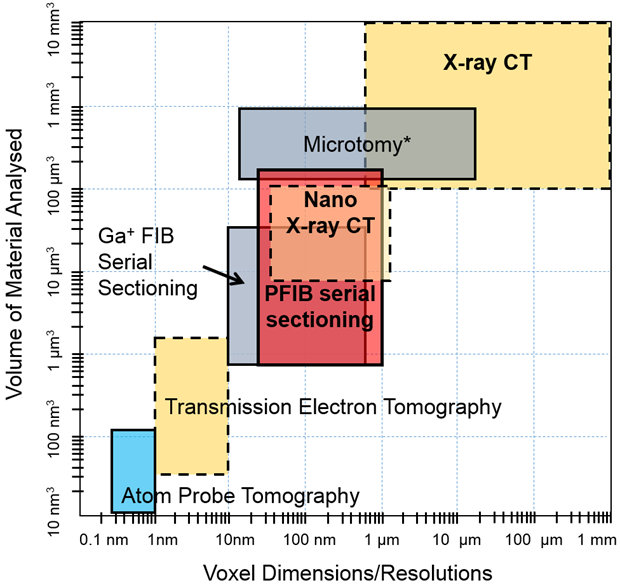
Recent advances in automated in situ SEM, dual beam and three beam scanning electron microscopy along with X-ray computed tomography (CT) have radically extended the range of scales that can be examined in 2D+time, 3D and 3D+time (see Fig. 1). Further, multi-modal correlative imaging can augment this with crystallographic and chemical information (see Fig. 2).
Figure 1: Through a combination of x-ray computed tomography and electron microscopy a very large range of scales can be probed for the same region of interest in a sample.
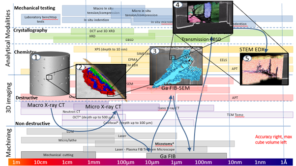
Through the digital image correlation of successive 2D or 3D images we can follow events as a function of growth, deformation or exposure to harsh environments with unprecedented precision. Further,by combining destructive and non-destructive techniques we can locate regions of interest in large volumes and then examine them in greater detail by combining different imaging modes to bring together complementary information (see Fig. 2).
Figure 2: Workflows can be established combining non destructive and destructive imaging to provide multiscale and multi-dimensional information – in this case to study pitting and intergranular corrosion in a steel.
Critical to the work flow is the use of new three beam (e.g. laser+Xe plasma FIB+electron) microscopes that enable regions of interest identified by CT to be examined at higher resolutions in 3D by electron microscopy.
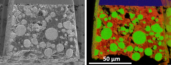
Figure 3: Triple beam (Laser PFIB) electron microscope slice of SE (left) and low kV EDS (right) to separate out pores and binder for a Lithium-Nickel-Manganese-Cobalt-Oxide (NMC) battery electrode.
Through a series of examples ranging from lithium battery electrodes (see Fig. 3), to butterfly pupation, to stress corrosion and from experimental steering to locate parasites in the gut, to mapping creep deformation at the grain scale in magnesium alloys, we will illustrate the opportunities and discuss the practical limitations of multiscale, multi-modal imaging.