Electron Microscopy Sample Preparation with Femto-second Laser Ablation
- Abstract number
- 488
- Corresponding Email
- [email protected]
- Session
- Correlative and Multimodal X-ray Microscopy
- Authors
- Dr. Andrew Elliott (1)
- Affiliations
-
1. ZEISS
- Keywords
femto-second laser, laser ablation, FIB-SEM, Focused ion beam scanning electron microscopes,
- Abstract text
Focused ion beam scanning electron microscopes (FIB-SEM) are highly versatile tools for the preparation, imaging and analysis of specimens. These tools have enabled researchers to create small samples, such as TEM lamellae, with greater ease, less time and reduced risk of sample loss compared to previous methods. However, the preparation of these microscale specimens if often the rate-limiting step in an analysis workflow. FIBs are excellent for precise machining but have limited milling rates, even with plasma-based sources. This limited milling rate means maximum volume that can be milled in a reasonable time to ~0.125 mm3, limiting the sample sizes/cross-sections that can be created. One answer to solve this is by utilising femto-second laser ablation as part of the FIB-SEM sample preparation workflow.
Laser ablation can be used to rapidly prepare large cross-sections, upto mm2, in a wide variety of materials. Such lasers can mill a mm3 in <<30 mins, a feat that would take a Ga+ FIB over 100 days. Until recently, the pulse width of laser ablation systems meant significant thermal energy is imparted to the milled surface. This creates a heat affected zone (HAZ) several microns thick, resulting in an altered microstructure. So, whilst previous nano-second laser ablation could mill rapidly, the resulting damage was too significant to be useful in most applications. But recent advances in laser technology have resulted in the advent of commercially available femto-second laser ablation solutions. The reduced pulse width results in athermal ablation with a significantly reduced damage zone, typically <0.5 microns. Surfaces produced by femto-second laser ablation are sufficiently smooth to be analysed directly with EBSD. If desired, the small sub-micron damage layer can be removed quickly with FIB.
This presentation will show the wide-ranging uses of femto-second laser ablation in preparation of EM samples; from large cross-sections to arrays of micromechanical testing specimens and even to uses in TEM lamellae preparation. Femto-second laser ablation reduces the time required to prepare samples, improving the time-to-results for users and resulting in higher system throughput.
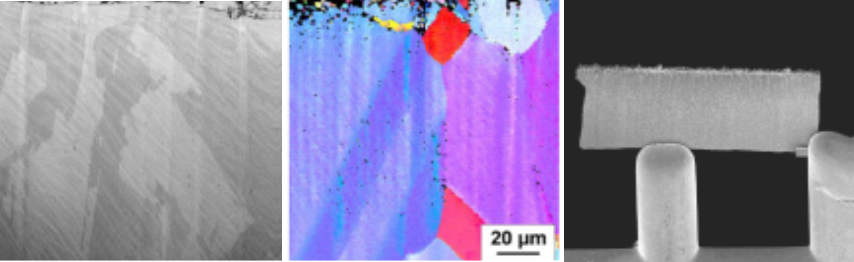
Figure 1 – Examples of laser ablated samples. Left) Cross-section in steel alloy 600 (image width ~120 μm). Middle) IPF map of Copper polished with with NOC filter. Right) Large TEM Lamella with standard Lamella for comparison