Fast and Deep 3D Live Cell Imaging
- Abstract number
- 487
- Corresponding Email
- [email protected]
- Session
- Optical Imaging of Fast Dynamic Processes
- Authors
- Mrs Karen McGeachy-West (1)
- Affiliations
-
1. Laser 2000 (UK) Ltd
- Keywords
Live Cell Imaging
confocal microscopy
Phototoxicity
- Abstract text
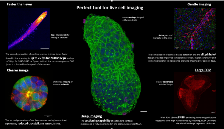
Phototoxicity is a central problem in the application of confocal microscopy for live cell imaging. To overcome these challenges, Laser 2000’s partner, Confocal.nl, developed the second generation of the fast line-scanning confocal technologies. Using a line scanner instead of a point scanner, the confocal microscope provides high temporal resolution, at the same time reducing the phototoxicity and allowing long-term live cell imaging. The new and improved version of this technology, NL5+, enables even faster imaging with cleaner results due to the additional motorized single-band emission filters.
The NL5+ takes the best of the two worlds, point scanners and spinning disc, leading to a system with options to do confocal, widefield and brightfield observations. NL5+ is optimized for fast and deep imaging with the lowest possible exposure of cells to the toxic effects of light.
Imaging information:
Calcium imaging in the root of A. thaliana. 7 days old root expressing nuclei targeted calcium sensor, treated with flg22 synthetic peptide. Maximum projection of 15 Z planes, with the total depth of 140 μm. Staining: Nuclei targeted green calcium sensor. Scale bar 200 μm. Acknowledgment to Dr. Mayuri Sadoine and Prof. Dr. Guido Grossmann, ICIB, Heinrich Heine University, Düsseldorf, Germany.
Left down – Selected single Z plane in mouse spheroid imaged in 60 μm depth. The difference between positive (green) and negative cells (blue) is clearly visible. Scale bar 20 μm. Sample courtesy of Nanami Satoh, MRC LMB.
Middle – Mouse embryo cultured in vitro for 24 h and imaged at 140 μm depth through 282 Z planes. Staining: DAPI, Phalloidin-488, HH3-647. Scale bar 20 μm. Sample courtesy of Nanami Satoh, MRC LMB.
Right up – Astrocytes and microglia in the neonatal mouse brain. Staining: GFAP-positive astrocytes (red), Iba-1 positive microglia (green), nuclei in DAPI (blue). Scale bar 30 μm. Sample courtesy of Dr. Eoin O'Neill from the Immune Regulation Research Group at Trinity College Dublin.
Right down – Mouse spinal cord. Synaptophysin labelled with Alexa555. Imaging with 561 nm laser followed by stitching. Scale bar 200 μm. Sample courtesy of Dr Matthew Broadhead, School of Psychology and Neuroscience, University of St Andrews.