Magnetic phase imaging using Lorentz near-field electron ptychography
- Abstract number
- 175
- Presentation Form
- Contributed Talk
- DOI
- 10.22443/rms.mmc2023.175
- Corresponding Email
- [email protected]
- Session
- EMAG - 4D STEM
- Authors
- Mr Shengbo You (3), Mr Penghan Lu (1), Dr Thomas Schachinger (2), Dr Freddrick Allars (3), Dr Andras Kovacs (1), Prof Rafal Dunin-Borkowski (1), Dr Andrew Maiden (3)
- Affiliations
-
1. Forschungszentrum Juelich
2. TU Wien
3. University of Sheffield
- Keywords
Lorentz mode, near field electron ptychography, phase imaging
- Abstract text
Ptychography is a phase-retrieval approach that has been widely used in electron microscopy society. It uses a localised ‘probe’ beam of illumination to scan through the sample with overlapping between adjacent scan positions. The intensity from recorded diffraction pattern is iteratively calculated to recover the complex transfer function of both sample and illumination probe. The redundancy introduced by the overlapping probes makes ptychography robust and insensitive to noise.
Over the past few years, near-field ptychography has drawn more attention for its ability to recover a large field of view with fewer diffraction patterns [1, 2]. Near-field ptychography replace the localised probe with a full-field structured illumination and move the detector from far-field to near-field. Recent research showed that by using a diffuser, the field of view can be as large as 100 , while only using a few diffraction patterns [3].
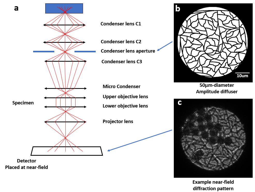
In this talk, we experimentally demonstrate that near-field electron ptychography can be used to reconstruct magnetic sample under Lorentz mode with various illumination sizes. We move the diffuser from selected area aperture to condenser lens. This improves the does efficiency and allow us to use different illumination sizes, while keeping the merits such as large field of view with few diffraction patterns. The purposed-designed amplitude diffuser will eliminate inelastic scattering and make the illumination more coherent. A parallel beam of electrons will illuminate onto the specimen. A system diagram is shown in Figure 1, together with a picture of the amplitude diffuser and an example of diffraction pattern. The reconstructed sample results are shown in Figure 2.
We would like to acknowledge the funding from European Union’s Horizon 2020 research and innovation programme under grant agreement No. 823717 (ESTEEM 3) and No. 856538 (“3D MAGiC”), and Royal Society International Exchange Award No. IESR2192202.
Figure 1: System diagram of near-field electron ptychography. The amplitude diffuser, shown in (b), is inserted at condenser lens aperture. An example of diffraction pattern is shown in (c), this diffraction pattern is under conventional TEM mode with latex sphere as sample. Under Lorentz mode, both upper and lower objective lens will be turned off due to the use of magnetic sample.
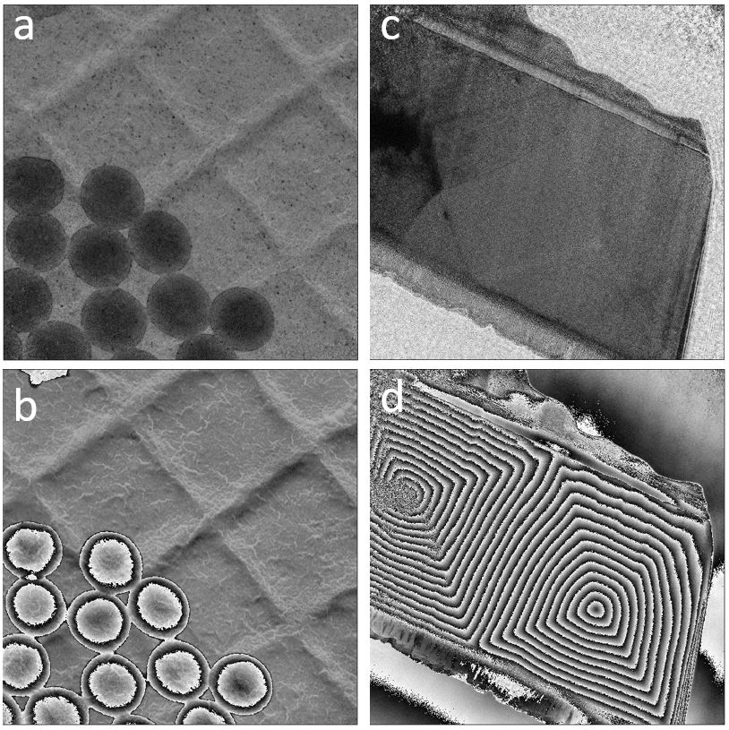
Figure 2. Latex sphere and Mo-doped Permalloy sampled reconstructed results under Lorentz mode. (a) and (b) are the reconstructed amplitude and phase of latex sphere sample. (c) and (d) are the reconstructed amplitude and phase of Mo-doped Permalloy. As for the magnetic sample (sub-figure c and d).
- References
[1]. M. Stockmar, et al, Sci. Rep. 3(1), (2013).
[2]. S. McDermott and A. Maiden, Opt. Express 26(19), 25471–25480 (2018).
[3]. F Allars and P Lu, Ultramicroscopy,231, (2021), p.113257.