Manipulation of the Point Spread Function of Light to Improve Quantum Limited Estimation Accuracy
- Abstract number
- 386
- DOI
- 10.22443/rms.mmc2023.386
- Corresponding Email
- [email protected]
- Authors
- Mr Joseph Baker (1)
- Affiliations
-
1. University of Birmingham
- Keywords
Information Theory
Bio-Medical Imaging
microscopy
- Abstract text
By including phase in a imaging system Rayleigh's curse is rendered irrelevant to our distance estimation problem. Work by Tsang showing that by including phase information in an imaging system then the amount of information extractable is unchanged, but this is cumbersome to implement for bio-imaging. Point spread function shaping, is an effective compromise. By using a one-dimensional dark ring one could improve estimation accuracy while being invariant to optical axis orientation.
Deterministic and Stochastic Super-Resolution techniques can regularly achieve resolutions well below 20nm [1]. However, this is only achieved via sophisticated control of the emission of fluorophores, and it is both time-consuming and invasive because super-resolution assumes individual emitters that can be resolved independently. There are nonetheless scenarios where one would like to localise simultaneously emitting fluorophores that are too closed to be individually resolved. This is in part due to progress made in the determination of the number of simultaneous emitters in unresolved clusters [2].
Here, we restrict ourselves to two identical but incoherent sources. In conventional intensity imaging, estimating the positions of two point-like sources becomes harder the closer these sources are to each other. Work by Tsang et al [3] showed that remarkably, this need not be the case: by capturing phase information in the imaging system, the amount of information on the separation of the sources that can be extracted from the radiation does not depend on the separation itself, provided the emitters are incoherent. Unfortunately, the corresponding implementations of such optimum scheme are cumbersome. They involve interferometric arrangements that are ill-suited to microscopy because the position of the centre of mass of the two sources would need to be known a priori, a rare occurrence in bio-imaging. As a practical compromise, Paúr et al [4] found that shaping the point spread function (PSF) of the imaging system such that it exhibits a zero of intensity, a comparatively easier operation, significantly improves the quantum-limited estimation of the separation. While the resolving power of this technique does still drop to zero at short distances, the scaling is much more favourable than imaging with a typical Gaussian point spread function.
While the original work of Paúr et al focused on a one-dimensional demonstration, we are interested in the two-dimensional case, where the direction of separation between the emitters is unknown a priori. By manipulating the phase of the radiated light such that the point spread function has a dark ring, we have shown analytically and numerically that the separation of two fluorophores can be estimate to an accuracy greater than direct imaging. A proof-of-principle experiment is under construction to confirm these predictions.
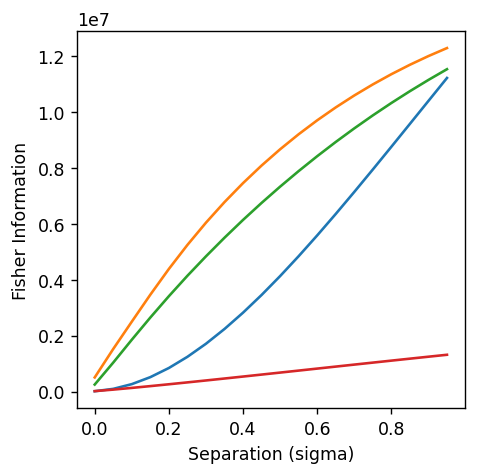
By manipulating the phase such that the point spread function has a dark ring we have shown analytically that the separation of two fluorophores can be estimate to an accuracy greater than direct imaging. This is quantified using the Fisher information – from which we can calculate the maximum amount of information theoretically extractable from a system, in this case two unresolved emitters shown in the Figure below. By virtue of using a one-dimensional ring no prior knowledge of the optical axis along which the fluorophores are separated is needed – a requirement for bio-medical imaging. By clipping a lens and adding aberrations we have generated a dark ring that achieves near extinction in the one-dimensional ring of zeros. By overlapping two 633nm He-Ne laser beams we can model two unresolved fluorophores and by separating them a known distance we can compare our imaging system with direct imaging.
Figure of numerical simulations showing that the dark ring (green) scales closely with the signum mask proposed by Paur and scales with separation better than direct imaging (blue) and the vortex plate (red)
This poster presents analytical derivations for the dark ring showing it’s preferable to other PSF shapes, numerical simulations accounting for Poisson and Gaussian noise and preliminary experimental results of our generated dark ring.
- References
[1] - Sahl, Steffen J., Stefan W. Hell, Stefan Jakobs. Fluorescence nanoscopy in cell biology. Nature reviews Molecular cell biology 18 11 685-701 (2017)
[2] - Hummert, Johan, Stanimir Asenov Tashev, and Dirk-Peter Herten. An update on molecular counting in fluorescence microscopy. The International Journal of Biochemistry & Cell Biology 135 (2021)
[3] - Tsang Mankei, Ranjith Nair, and Xiao-Ming Lu. Quantum theory of super resolution for two incoherent optical point sources. Physical Review X 6.3 (2016)
[4] - Paúr, Martin, et al. Tempering Rayleigh’s curse with PSF shaping. Optica 5.10 (2018)