Nanomechanical analysis with photothermally excited cantilevers.
- Abstract number
- 554
- Corresponding Email
- [email protected]
- Session
- AFM in Life Sciences
- Authors
- Dr Héctor Corte-León (1), Mr Hans Gunstheimer (1), Mr Gotthold Fläschner (1), Dr Jonathan Adams (1), Dr Pieter van Schendel (1)
- Affiliations
-
1. Nanosurf
- Keywords
AFM life-sciences bio nanomechanics photothermal nanoscale
- Abstract text
Atomic force microscopes not only provide morphological information, but mechanical information about the sample under study can be obtained as well. This is of particular interest in the biophysical sciences, because this process can be done at ambient pressure, in both air and liquid.
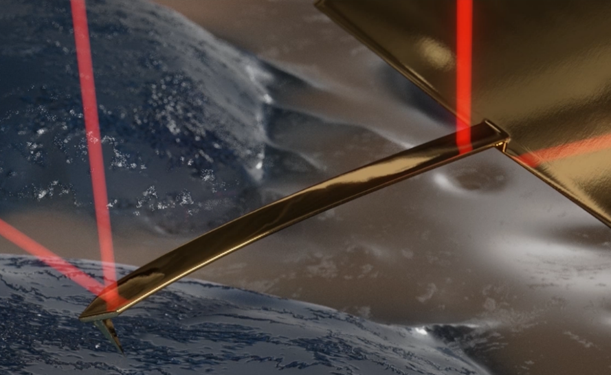
To measure mechanical properties, the AFM system requires performing a force vs distance measurement at every single point of the area of interest. Typically, with piezo-actuated AFM systems, this process is limited by the bandwidth of the actuator, and additionally the stability of the medium when in liquid. With photothermal excitation off-resonance tapping (Fig. 1), these limits are overcome. The photothermal excitation is used to drive the AFM cantilever up and down. In this way, the limit of the piezo bandwidth is lifted, and the part of the system that oscillates is significantly smaller, and the data acquisition speed is eventually only limited by the AFM probe bandwidth and data acquisition rate. In liquid the direct cantilever driving is beneficial because it reduces noise caused by the liquid movement, which is larger in piezo actuated systems.
Figure 1: schematic representation of using photothermal actuation to directly actuate cantilever bending, through a second, intensity-modulated light source that is directed towards the base of the cantilever.
In addition photothermal excitation can be used to dynamically probe the viscoelastic properties of samples at a large range of frequencies.
In summary , photothermal excitation enables fast, stable mechanical sample characterization, in both air and liquid.
- References
[1] A.P. Nievergelt, et al., Nature Nanotech 13, 696–701. (2018)