Native state structural and chemical characterisation of Pickering emulsions: A cryo-electron microscopy study
- Abstract number
- 333
- Presentation Form
- Contributed Talk
- DOI
- 10.22443/rms.mmc2023.333
- Corresponding Email
- [email protected]
- Session
- EMAG - Bio, Cryo & Low-dose EM Imaging
- Authors
- Dario Luis Fernandez Ainaga (1), Martha Ilett (1), Teresa Roncal-Herrero (1), Stuart Micklethwaite (1), Cheng Cheng (1), Olivier Cayre (1), Nicole Hondow (1)
- Affiliations
-
1. University of Leeds
- Keywords
Pickering emulsion
Cryo-TEM
Cryo-FIB-SEM
EELS
EDX
Electron Tomography
- Abstract text
Summary –
Cryo-electron microscopy offers huge potential in the imaging and analysis of aqueous inorganic samples if appropriate measures are taken in limiting electron beam damage and subsequent devitrification of the ice and sample. In this study, an oil-in-water Pickering emulsion stabilised by platinum nanoparticles was plunge-frozen and successfully imaged in cryo-TEM using cryo-STEM, EELS, EDX, and cryo-tomography. A sample lamella prepared in cryo-FIB-SEM was cryo-transferred to a TEM for the imaging of emulsion samples with larger droplet sizes, otherwise unsuitable for plunge-freezing.
Introduction -
Cryogenic electron microscopy (Cryo-EM) permits imaging of aqueous samples in the vacuum of an electron microscope while preserving the structure of the sample. Although cryo-EM is extensively and routinely used for the imaging of biological samples (e.g. cells, viruses, tissue samples), it isn’t as widely applied to inorganic samples.
This cryo-EM study examines an inorganic sample, an oil-in-water Pickering emulsion stabilised by platinum nanoparticles. These emulsions can have applications in drug delivery, chemical and food products, and therefore their accurate characterisation is required understanding of their stability and performance. The droplets and stabilising nanoparticles provide both large (50 nm – 10 mm) and small (~5 nm) scale objects that need a combination of low-magnification and high-resolution imaging to enable accurate characterisation. Utilising instrumentation designed for the physical and materials sciences, the elemental analysis techniques electron energy loss spectroscopy (EELS) and energy dispersive X-ray (EDX) spectroscopy have been used to further examine the Pickering emulsion sample.
In this research, Pickering emulsions were cryo-prepared for a range of EM imaging and analysis approaches while maintaining a vitrified sample to better understand how to adapt characterisation techniques widely used for dry sample imaging to cryogenically prepared samples.
Methods/Materials -
Hexadecane in water Pickering emulsions stabilised with ~5nm platinum nanoparticles were plunge-frozen in liquid ethane for cryo-TEM imaging in a FEI Titan Themis3 TEM at 300kV. EDX and EELS data was collected in cryo-STEM using a Super-X 4-detector silicon drift energy dispersive X-ray system and a Gatan GIF Quantum 965 electron energy loss spectrometer. Tilt series for tomography reconstruction were taken in cryo-STEM using the FEI Tomography STEM software and reconstructed using SIRT in Inspect3D. Samples for cryo-FIB-SEM imaging were prepared by freezing the sample in liquid nitrogen and using a Quorum SEM cryo-transfer unit was transferred to a Helios G4 CX FIB-SEM.
Results and Discussion -
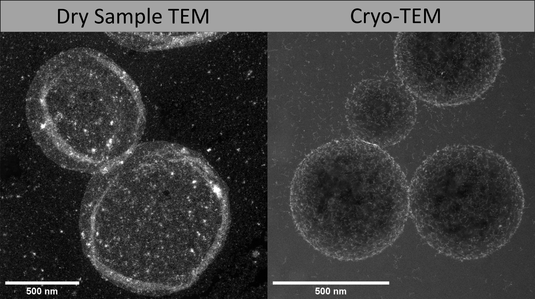
This work will show the advantage of using a cryogenic approach is evident when compared to images collected from a dried sample. In the dried sample the integrity of the emulsion droplets is not maintained, with most appearing to have burst when drying or due to the vacuum of the microscope. Meanwhile, cryo-STEM shows the emulsion droplets to be spherical with the size and shape as anticipated from bulk measures. Droplet shape was further been investigated using cryo-STEM tomography, where multiple images are collected at a range of sample tilt angles and then reconstructed to form a 3D image, confirming that the 3D spherical shape of the droplets is still present after sample preparation.
Cryo-STEM was preferred for sample imaging as compared to cryo-TEM due to the higher total electron fluence needed before incurring sample damage. In cryo-STEM it was possible to both image the large-scale emulsion droplets (diameter >100nm) and the stabilising Pt nanoparticles (~5nm), though STEM imaging, however when pixel sizes below 3nm x 3nm were used there was increased beam-induce damage preventing data collection prior to ice and sample damage. Following these electron dose specifications, cryo-EDX and cryo-EELS were successfully used to map the presence of carbon in the oil phase of the emulsion. Cryo-electron tomography was also employed to image the sample in 3D and to obtain a 3D elemental map using cryo-EELS while simultaneously obtaining a cryo-STEM tilt series.
Despite the successful imaging of the plunge-frozen sample, the thickness requirements of cryo-TEM imaging limits the range of sample sizes for this technique. To remedy this, a 300 nm lamella of a sample with >1 mm sized emulsion droplets was prepared in a cryo-FIB-SEM and then transferred for cryo-TEM imaging. This method allowed for higher-resolution imaging of the sample, therefore obtaining more information on the position and size of the stabilising nanoparticles.
Conclusions -
Cryo-EM was successfully used to image plunge-frozen Pickering emulsion samples using cryo-STEM, -EELS and -EDX. When attempting to analyse cryogenically prepared samples, beam-induced damage becomes the largest barrier for obtaining high-magnification images and high signal-to-noise ratios. Despite this, representative characterisation of inorganic samples in cryo-TEM is possible following the determination of electron dose limitations.
Additionally, there are still concerns regarding the reliability of plunge-freezing to maintain the structure and size of droplet clusters in their native state. Sample preparation in a cryo-FIB-SEM and transferring a lamella for cryo-TEM imaging can help mitigate these sample preparation challenges while also allowing for the imaging of cross-sections of larger samples.
- References

