Near-field Multislice Ptychography - Demonstration Using Visible Light and X-ray experiment
- Abstract number
- 149
- Presentation Form
- Contributed Talk
- DOI
- 10.22443/rms.mmc2023.149
- Corresponding Email
- [email protected]
- Session
- Correlative and Multimodal X-ray Microscopy
- Authors
- Mr Ziyang Hu (2), Mr Yiqian Zhang (2), Dr Peng Li (1), Dr Darren Batey (1), Dr Andrew Maiden (2, 1)
- Affiliations
-
1. Diamond Light Source
2. University of Sheffield
- Keywords
Quantitative Phase Image; Ptychography; Multislice; X-ray imaging; imaging system;
- Abstract text
Ptychography is a lensless coherent diffractive imaging technique. It has shown the potential in pushing resolution limitation in recent research across all wavelength regimes and ptychography is commonly applied for electron microscopy and synchrotron facilities[1]. However, it still remains a challenge to achieve such resolution for samples with a larger volume and the reconstruction process is excessively computational intensive. In this work, the authors demonstrate the implementation of near-field multislice ptychography (NMP) and show the experimental results in both visible light and x-ray wavelength.
Multislice was originally proposed by Cowley and Moodie, which is initially used to simulate the forward model of the multi-scattering process in transmission electron microscopy[2]. In this case, the multi-scattering process is replaced by multiple single scattering processes between each two sequential layers. However, the inverse model still remains a tricky problem and it is usually very difficult to find an accurate solution. In general, ptychography method requires to collect diffraction patterns from a series of overlapping illumination, then the iterative algorithm is fed with the redundant information to achieve the phase image reconstruction[3]. The diverse and redundant ptychographic data has proved advantageous and is able to solve the multiple 2D slices within the object. Therefore, we can establish the backpropagation model using ptychographic method. The reverse calculation can be derived from the object and probe updates in each layer. So far, multi-slice ptychography has been commonly used for the far-field experiments across all wavelengths. However, near-field ptychography shows advantage in obtaining a large field of view with the need of very few diffraction patterns and it has low requirement in detector dynamic range. Differently to the original 3PIE algorithm for far-field regime[4], we adapted the algorithm for near-field configuration using cone beam.
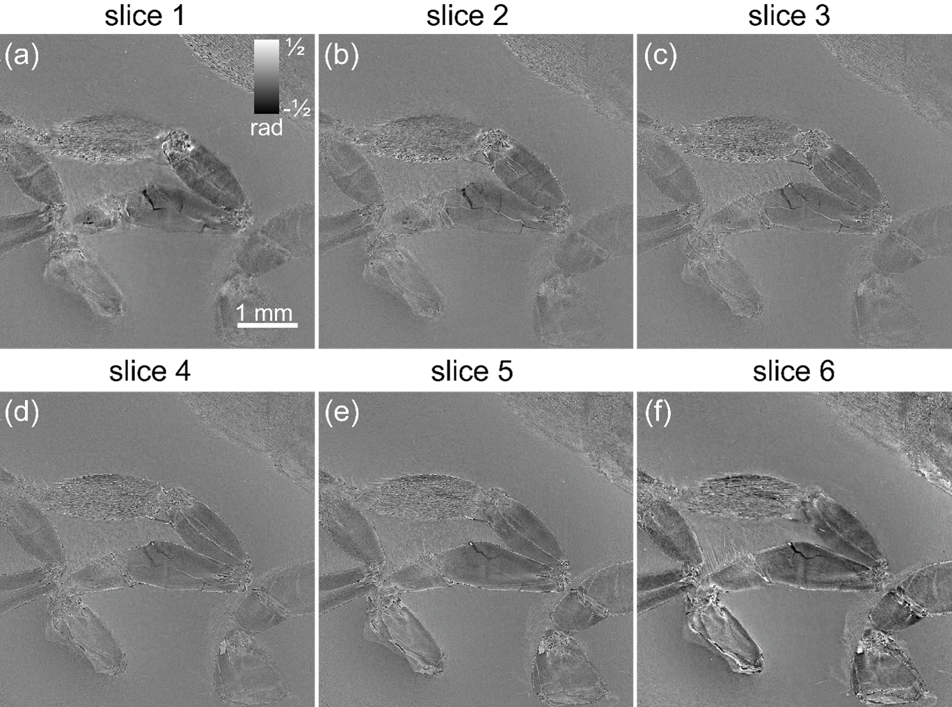
We firstly demonstrated the NMP result with visible light (675 nm laser) experiment. We achieved the multi-slice reconstruction of the phase image with sample thickness over 250 um and the field of view over of 5mm2 using only 15 diffraction patterns. The corresponding 6-layer reconstruction with 100 diffraction patterns is shown Fig.1(a)-(f), where a) is the most upstream layer (furthest to the detector) and f) is the most downstream layer (closest to the detector).
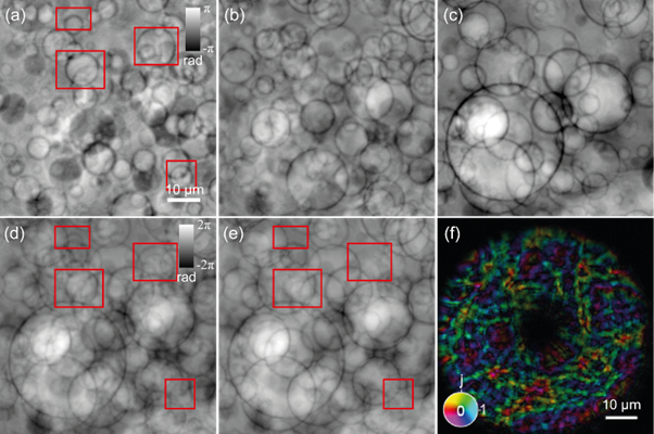
The x-ray multislice near-field ptychography was conducted in beamline I13-1, the Diamond Light Source, UK. A sheet of paper was inserted just upstream of the FZP to create a structured illumination.Under this experiment setup, the measured resolution is around 150-200 nm, and the theoretical z-axial slice distance is set to just over 0.5 mm. Fig. 2 shows the result of a 3-layer reconstruction with 1 mm separation distance of a continuous sample, which is a 2.5 mm thick paraffin block scattered with micro-sized glass beads. (a)-(c) corresponding to up to down beam layers. (d) is the pixel wise added result from 3 separated slices, (e) conventional 2D reconstruction and (f) probe reconstruction from 3PIE. The data set consists of diffraction patterns collected from 11 by 11 raster scan with 90% overlap. The reconstruction takes in total 500 iterations of 3PIE algorithm. The multislice reconstruction results show a significant improvement, in comparison to the conventional 2D ptychography reconstruction, which is limited by the DoF (Depth of Field). The features located beyond the DoF range can not be observed using ePIE reconstruction, as shown in the red box areas in Fig.2(a). The overall image quality, such as sharpness and contrast, also improves using the multislice method. We also further evaluated the minimal requirement of the diffraction pattern number from the same data set. We are still able to extract most of the features when it is reduced to 36 diffraction patterns and when the number is reduced to 20s’ the reconstruction starts to fail.
In conclusion, we can say that the multislice method is more beneficial for imaging samples, whose thickness exceeds the DoF limit for conventional 2D ptychography. NMP can address large sample volume while keeps the benefit of high resolution. Furthermore, we redefined the estimation of minimal slice separation distance by including the paraxial effect appeared in the experimental setup. The full potential of this technique will be revealed when it is combined with tomography.
Fig. 1. 6-layer multislice reconstruction of bee's legs with 10 by 10 diffraction patterns. (a) the most upstream layer b)-e) corresponding layers in between (f) the most downstream layer
fig. 2. 3-layer reconstruction of paraffin embedded glass beads with 11 by 11 diffraction patterns. (a) up-stream layer; (b) mid layer; (c) down-stream layer; (d) pixel-summed result from (a)-(c); (e) ePIE reconstruction; (f) reconstructed probe using multislice method.
- References
- F. Pfeiffer, “X-ray ptychography,” Nature Photonics, vol. 12, pp. 9-17, 2018.
- J Cowley, “Diffraction Physics,” 3rd Ed. North Holland Publishing Company, 1995
- J. Rodenburg and A. Maiden, “Ptychography,” in Springer Handbook of Microscopy, Switzerland, Spinger, 2019, p. 819.
- M. Maiden, M. J. Humphry and J. M. Rodenburg, “Ptychographic transmission microscopy in three dimensions using a multi-slice approach,” Journal of the Optical Society of America A, vol. 29, no. 8, pp. 1606-1614, 2012.