Chemical mapping of organic and bio materials with 10 nm spatial resolution using scattering-type scanning near-field optical microscopy (s-SNOM)
- Abstract number
- 80
- Presentation Form
- Poster
- Corresponding Email
- [email protected]
- Session
- Poster Session Two
- Authors
- Dr. Lars Mester (1), Dr. Philip Schäfer (1), Dr. Adrian Cernescu (1), Dr. Andreas J. Huber (1)
- Affiliations
-
1. attocube systems AG (neaspec)
- Keywords
Nanoscale imaging
Infrared nano-spectroscopy
Biological material
Scanning probe microscopy
- Abstract text
Scattering-type Scanning Near-field Optical Microscopy (s-SNOM) is a scanning probe approach to optical microscopy and spectroscopy, bypassing the ubiquitous diffraction limit of light to achieve a spatial resolution below 20 nanometers. s-SNOM employs the strong confinement of light at the apex of a sharp metallic atomic force microscopy (AFM) tip to create a nanoscale optical hot-spot. Analyzing the scattered light from the tip enables the extraction of the optical properties (e.g. absorption, reflection) of the sample directly below the tip and yields nanoscale resolved images simultaneous to topography. In addition, the technology has been advanced to enable Fourier-Transform Infrared Spectroscopy on the nanoscale (nano-FTIR) using broadband radiation from the visible and infrared spectral ranges to THz frequencies.
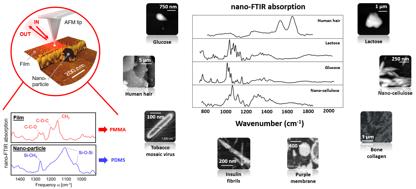
Figure 1 – Overview of nano-infrared studies on biomaterials using s-SNOM / nano-FTIR. Left: Illustration of nano-FTIR experiment and chemical identification of a nanoscale contaminant. Right: Nano-FTIR spectra of various biological samples.
Recently, the analysis of complex biological materials via correlative nano-optical and nano-mechanical imaging has gained significant interest. Nano-FTIR spectroscopy enables not only nano-chemical identification and to explore local distribution of organic and inorganic materials (Figure 1), but it also offers information about the secondary structure of proteins. Furthermore, nano-FTIR on a single ferritin complex even proofs sensitivity to about 4,000 amino acids, corresponding to about 5,000 C=O and N-H bonds, respectively.
Here we present various examples of s-SNOM measurements on different biological samples, demonstrating nano-chemical mapping of a-helices and b-sheets, as well as recent results on aggregated amyloid fibers relevant to neurodegenerative Alzheimer’s disease. Lastly, novel and unique instrumentation capabilities will be introduced, that extend the capabilities of commercial s-SNOM systems to being combined with fluorescence imaging and liquid sample environments.