Effect of hydrophobic and hydrophilic surfaces on the morphological and physical properties of supported lipid membranes
- Abstract number
- 150
- Presentation Form
- Poster & Flash Talk
- DOI
- 10.22443/rms.mmc2023.150
- Corresponding Email
- [email protected]
- Session
- Nanoscale Probing of Physical Properties via AFM & SPM
- Authors
- Harriet Read (1, 2), Dr Simone Benaglia (1, 2), Dr Laura Fumagalli (1, 2)
- Affiliations
-
1. University of Manchester
2. National Graphene Institute
- Keywords
Lipids, Bio-membranes, Atomic Force Microscopy (AFM), vdW crystals
- Abstract text
Lipid bilayers form the major component of the cell membrane, where the structure and properties of the membrane determine the functions that each cell is responsible for. Whilst supported lipid bilayers (SLBs) have long been accepted as basic model systems to study the cell membrane [1] , atomic force microscopy (AFM) has proved to be an effective tool to characterise the physical and thermodynamic properties of SLBs [2]. Among the different thermodynamic properties defining the SLBs, the lipid phase transition temperature, Tm, has been investigated extensively, with the transition from solid-ordered to liquid-disordered states known to play a substantial role into events in the cell such as membrane protein mobility [3, 4]. Studies regarding the phase transition of SLBs on hydrophilic substrates have been plenty, however, knowledge regarding the impact of hydrophobic substrates, such as van der Waals (vdW) crystals, on lipid phase transition is still lacking, which is of particular importance in the development of new bio- and electrochemical sensing technologies where conductive vdW crystal are used [5]. Additionally, it is expected that changes in thermodynamic properties of SLBs affect other physical properties such as their interfacial, conductive and dielectric polarisation properties.
Here, we present the study of supported phosphatidylcholine lipid membranes on both hydrophilic and hydrophobic substrates, where the morphological and thermodynamic properties of the membrane were investigated using temperature-controlled AFM. First, the morphological characteristics of lipid membranes on hydrophobic surfaces (forming stable lipid bilayers) differ from those on hydrophilic substrates (lipid monolayers with temperature independent ripple domains). Furthermore, the thermodynamic properties such as the transition temperature, enthalpy and the interaction between the lipid molecules during the transition vary greatly dependent upon the hydrophobicity of the substrate, with Tm of the transition increasing for hydrophobic substrates. We demonstrated the variation in thermodynamic properties affected similarly different phosphatidylcholine lipid, hence it is independent of the length of the lipid tail. Finally, by using different hydrophobic substrate, i.e. HOPG and hBN, we attempted to determine whether the change in physical properties were due to the hydrophobicity of the surface, or rather the difference in surface composition of the substrate.
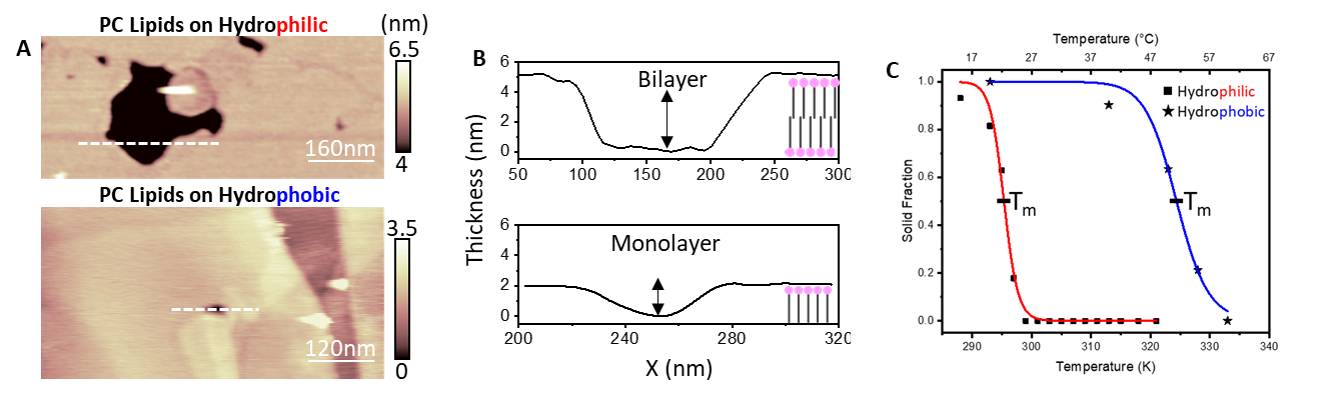
Figure 1: A) Morphology of PC-lipid bilayers and monolayers on hydrophilic and hydrophobic surfaces respectively. B) Associated profiles and configuration of lipids. C) The fraction of the membrane in the gel form with respect to temperature – the transition temperature may be extracted, showing a higher Tm for hydrophobic surfaces.
- References
[1] E. Sackmann, “Supported Membranes: Scientific and Practical Applications,” Science, vol. 271, pp. 43-48, 1996.
[2] M. Mingeot-Leclercq, M. Deleu, R. Brasseur and Y. Dufrene, “Atomic force microscopy of supported lipid bilayers,” Nature Protocols, vol. 3, pp. 1654-1659, 2008.
[3] A. Alessandrini and P. Facci, “Phase transitions in supported lipid bilayers studied by AFM,” Soft Matter, vol. 10, pp. 7145-7164, 2014.
[4] Y. Lin, C. Chipot and S. Scheuring, “Annexin-V stabilises membrane defects by inducing lipid phase transitions,” Nature Communications, vol. 11, p. 230, 2020.
[5] H. Bi, X. Wang, X. Han and K. Voitchovsky, “Impact of electric fields on the nanoscale behaviour of lipid monolayers at the surface of graphite in solution,” Langmuir, vol. 34, pp. 9561-9571, 2018.