Continuous Multiple Pass Electron Counted Spectrum Imaging for High Temporal Resolution In-Situ Analysis
- Abstract number
- 574
- Presentation Form
- Poster
- Corresponding Email
- [email protected]
- Session
- Poster Session Three
- Authors
- Dr Liam Spillane (2), Dr Benjamin Miller (2), Dr Anahita Pakzad (2), Dr Michele Conroy (1)
- Affiliations
-
1. Department of Materials, Imperial College London
2. Gatan
- Keywords
- Spectroscopy and spectrum imaging
- In situ and dynamic microscopy techniques and development
- Functional semiconductor, oxide and advanced materials
- Abstract text
Continuous zero dead time multiple pass scan capabilities of a flexible next generation scan system (Digiscan3) are deployed to explore the utility of single electron counting direct detection cameras for in-situ spectrum image (SI) acquisition at high temporal resolution.
For in-situ analysis, the ability to capture data at high speed is critical. For SI data capture this requirement must be fulfilled by both the STEM scan system and the detector used for data capture. A prototype approach for In-situ SI acquisition was recently demonstrated using a current generation CMOS based electron energy-loss spectroscopy (EELS) spectrometer (GIF Continuum) and Gatan DigitalMicrograph [1]. Copper (II) oxide was subjected to in-situheating using a MEMS based heating holder (Wildfire, DENSSolutions) to test this new paradigm. Energy-loss near-edge structure (ELNES) mapping performed on Cu L2,3 spectra both live and in post analysis showed a change in Cu oxidation state from majority Cu(II) at low temperature to majority Cu(0) at elevated temperature. Spontaneous changes in particle morphology were also observed at specific temperatures. Whilst these results clearly validated the in-situ SI acquisition modality, limitations were also observed. Non-zero dead time between SI acquisition passes was found to be a major fraction of total capture time for very short SI pass time. Detector read noise associated with indirect detection also constrained the electron dose rate that could be used to acquire high signal-to-noise data and therefore camera frame rates that were practical to use.
Single electron counting direct detection cameras are optimal for in-situ analysis due to their intrinsic high detective quantum efficiency (DQE) and high frame rate readout capability. As read noise is eliminated by electron counting, it is typically possible to maximise temporal resolution by running at the maximum camera frame rate [2]. In-situ frames can then be combined as required to balance signal-to-noise against temporal resolution during post acquisition data analysis. By using such detectors with a flexible next generation scan control system (Digiscan3) it has been possible to perform continuous multiple pass SI acquisition over large rectangular arrays at sub-second total pass time. This significant improvement in temporal resolution is achieved due to zero-dead time multiple pass scan and hardware synchronized sub-pixel scanning capabilities of the new scan control system. This high temporal resolution scan setup is ideal for in-situ analysis.
In this work, a number of different materials systems have been subjected to in-situ heating to explore the benefits ofenhanced temporal resolution SI acquisition. These materials include: metal nanostructures and oxide powders such as Fe2O3.H2O as shown in figure 1. Temporal resolution and heating ramp rate was systematically varied to determine the need for synchronized holder control. To explore the requirements for multi stimuli in-situ experiments, reversible phase changes in multiferroic boracite (M₃B₇O₁₃X) domains and domain walls were investigated by in-situ heating, cryogenic cooling and biasing. The dynamics of the domain walls is directly related to the changes in the local wall symmetry, and in the case of the transition metal-based boracites the ability of the oxidation state to easily and reversibly switch between states. Thus, it is essential to do in-situ EELS L2,3 fine structure analysis of these elements during domain wall dynamics. The transition metal (Cu, Fe and Co) L2,3 fine structure changes were analysed during cycling of ferroic phases with corresponding atomic scale STEM imaging, thus relating the evolving physical distortions of the unit cell to the spectra. A discussion of experimental design considerations for single and multi stimuli in-situ spectrum image are is then presented.
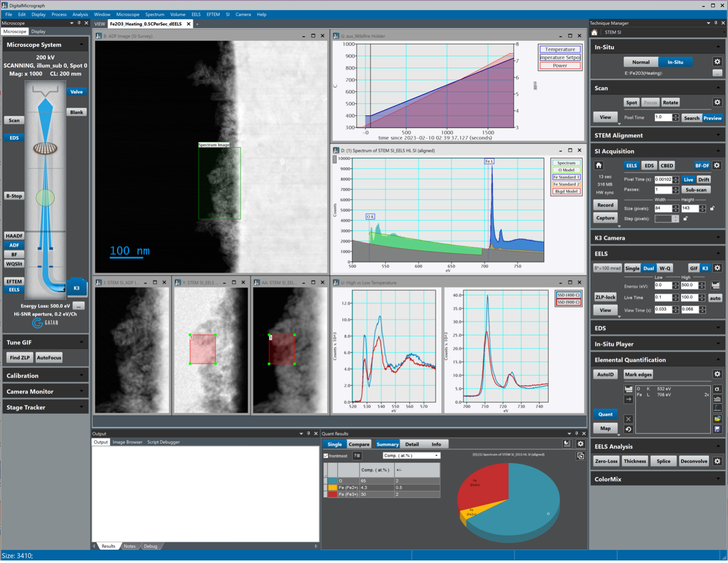
Figure 1. DigitalMicrograph screenshot showing in-situ EELS spectrum image dataset acquired during in-situ heating performed on Fe2O3.H2O from 400°C – 900°C with ramp rate 0.5°C/s. Data was acquired in dualEELS mode using a single electron-counting camera (Gatan, K3). Live quantification using concurrent standards acquired from FeO and Fe2O3 references showed a transition from majority Fe(III) to Fe(II) at elevated temperature.
- References
[1] Spillane, L. et al., Microsc. Microanal. 26 (Suppl 2), 2020, p.1676
[2] Hart, J. L., et al., Direct Detection Electron Energy-Loss Spectroscopy: A Method to Push the Limits of Resolution and Sensitivity. Sci Rep 7 (2017): 8243.