Phase contrast from 4D-STEM data at finite electron dose
- Abstract number
- 316
- Presentation Form
- Contributed Talk
- DOI
- 10.22443/rms.mmc2023.316
- Corresponding Email
- [email protected]
- Session
- EMAG - 4D STEM
- Authors
- Dr Laura Clark (3), Dr Scott Findlay (1), Prof Pete Nellist (2)
- Affiliations
-
1. Monash University
2. University of Oxford
3. University of York
- Keywords
4D-STEM, phase contrast, ptychography, DPC STEM, low dose, beam sensitive
- Abstract text
In recent years, we’ve seen both increasing use of phase-imaging methods based on pixelated detectors (as highlighted in EMAG 2020) and increasing awareness of the need to use restricted electron dose when imaging some materials (as highlighted in EMAG 2022). Here, we discuss how the former methods are limited by the latter noise problems.
Pixelated detectors are increasingly widely available – at both local and national facilities. 4D-STEM imaging methods are starting to be used by a broader community, and for increasingly challenging specimens (that is – in cases where the noise properties of the image are non-negligible) . When considering the effect of noise on the resultant image, we must recall that 4D-STEM imaging methods are fundamentally different to conventional (S)TEM imaging methods, in that conventional methods form direct images without additional processing and the image noise will be Poisson-dominated (in a well-behaved microscope – such that effects like drift and vibrations are negligible) [1]. 4D-STEM methods, however, are computational imaging techniques, in that the recorded data is processed through some algorithm, prior to forming the image [2]. The noise in the recorded data is thus also processed through the same algorithm, which impacts how it appears in the final image.
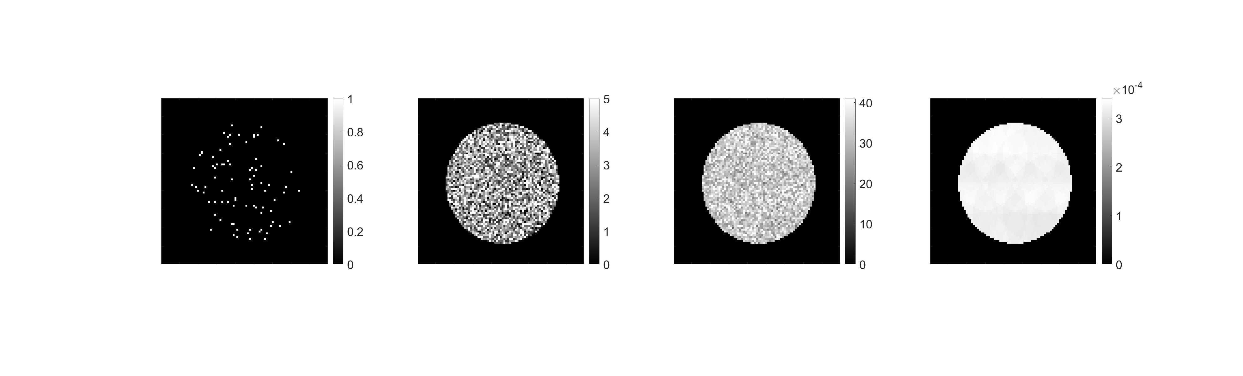
More specifically, we look at the limits achievable for different 4D-STEM imaging methods (i.e., differential phase contrast (DPC), centre of mass (CoM ,) and ptychography) at different dose levels and imaging settings (following Refs [3,4]). We discuss the non-trivial correlated noise that appears in images computed from 4D-STEM datasets, the smoothing versus coarsening effects of the different imaging processes, and connections to prior work on conventional TEM imaging processes [5] and 4D-STEM imaging of thick samples [6].Figure 1: Simulated brightfield discs (input to 4D-STEM analyses) at varying electron dose levels: 100, 10,000, 100,000 and infinite dose. The fine-grained details key to precise interpretation form only slowly with increasing dose.
LC acknowledges funding from a Royal Society University Research Fellowship (URF\R1\221270) and additional Royal Society funding (RF\ERE\221035). This research was partly supported under the Discovery Projects funding scheme of the Australian Research Council (Project No. FT190100619).- References
[1] Chen, Z, et al. Nature Communications 11.1 (2020): 2994.
[2] Gureyev, TE, et al. Physical Review A 97.5 (2018): 053819.
[3] O’Leary, CM, et al. Ultramicroscopy 221 (2021): 113189.
[4] Seki, T, et al. Ultramicroscopy 193 (2018): 118-125.
[5] Clark, L, et al. Micron 124 (2019): 102701.
[6] Clark, L, et al. Microscopy and Microanalysis 29.1 (2023): 384-394.