Quantitative analysis of atomic displacements in iron/iron oxide core-shell nanoparticles and their effect on high-resolution scanning transmission electron microscopy imaging
- Abstract number
- 359
- Presentation Form
- Contributed Talk
- DOI
- 10.22443/rms.mmc2023.359
- Corresponding Email
- [email protected]
- Session
- EMAG - High Resolution Structural Analysis
- Authors
- Shuayl Alotaibi (1, 2), Demie Kepaptsoglou (1, 3), Andrew Pratt (1), Roland Kröger (1)
- Affiliations
-
1. Department of Physics, University of York
2. Physics Department, Prince Sattam bin Abdulaziz University
3. SuperSTEM
- Keywords
Strain, displacement, STEM image, image intensity
- Abstract text
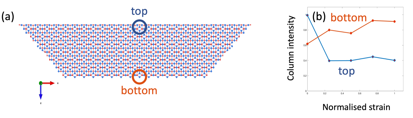
Strain in nanoparticles (NPs) plays a key role in controlling catalytic or chemical activity and its impact on the atomic level structure is essential for applications of such NPs in a wide variety of fields ranging from biomedicine to environmental remediation. Therefore, atomic level displacement analysis is a highly relevant research topic with characterisation techniques such as high-resolution scanning transmission electron microscopy (HRSTEM) to be employed in this context [1]. A fundamental challenge of analysing atomic level strain with HRSTEM in NPs is the impact of strain on the image formation since strain induces modifications of column intensities and intensity distributions and hence affects the interpretation of image contrast and column positions. To systematically study this impact, we used aberration corrected HRSTEM to investigate cluster-source-deposited partially oxidised cubic iron NPs and developed a systematic model of the magnetite oxide shell including grain boundary induced lattice strain. We created an atomistic model of the oxide shell, applied a finite element calculated strain field and simulated HRSTEM images of such strained domains using QSTEM [2] including thermal diffuse scattering. This model was inspired by our previous work indicating that lattice strain is induced by stress at the oxide domain boundaries [1]. Our data reveal that lattice strain has various effects on the HRSTEM images including a reversal of Fe(II) column intensity distribution within the domains in comparison to the unstrained case as seen in Fig. 1 as well as streaking due to strain-induced atomic displacements, which is in-line with experimental observations.
Fig. 1: (a) Unstrained model structure of magnetite domain. (b) top and bottom column intensities as a function of normalized strain.
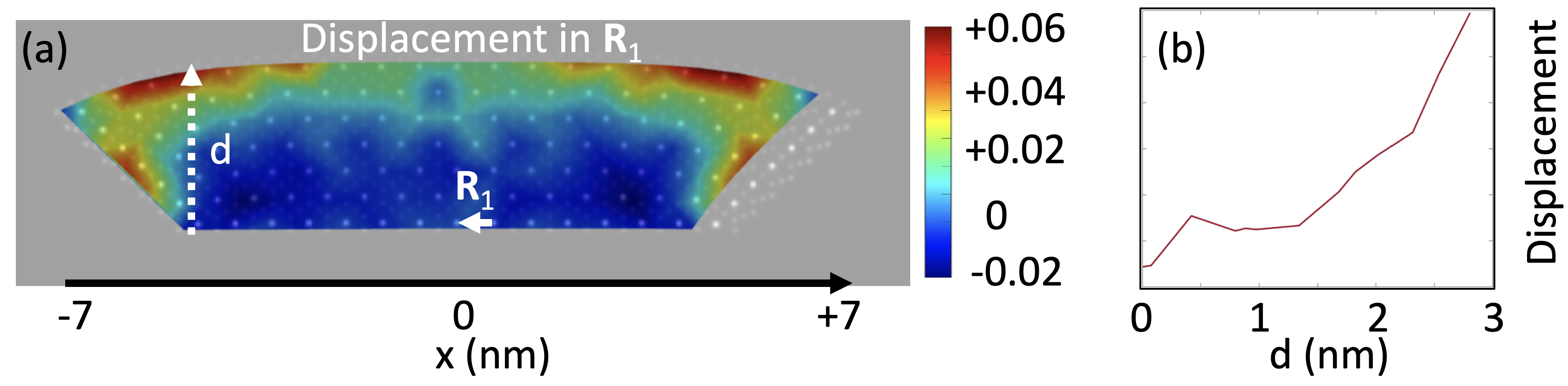
We further compared the atomic displacement fields derived from experimental images with those resulting from our image simulations (see. Fig. 2) showing a significant maximum displacement at the oxide surfaces and a linear strain gradient between the core/oxide boundary and the oxide surface, which is also in good agreement with experiment.
Fig. 2: (a) Overlay of simulated HRSTEM image including strain and the resulting atomic displacement map for lattice vector R1. (b) displacement profile along direction given by d in (a).
Overall, we find that atomic level displacements due to strain fields strongly affect HRSTEM image formation and need to be carefully considered when quantitatively analysing such data e.g. for the extraction of strain field information.
- References
[1] A. Pratt, L. Lari, O. Hovorka, A. Shah, C. Woffinden, S.P. Tear, C. Binns, R. Kröger, Enhanced oxidation of nanoparticles through strain-mediated ionic transport, Nat. Mat. 13, 26 (2014).
[2] C. Koch, Determination of Core Structure Periodicity and point defect density along dislocations, ProQuestDissertationsandTheses, Arizona State University (2002).