The ultrastructure of bone in 3D: A twist of twists
- Abstract number
- 319
- DOI
- 10.22443/rms.mmc2023.319
- Corresponding Email
- [email protected]
- Session
- FIB Applications & EM Samples Prep Techniques in Physical Sciences
- Authors
- Daniel Buss (1), Roland Kroeger (2), Marc McKee (1), Natalie Reznikov (1)
- Affiliations
-
1. McGill University
2. University of York
- Keywords
bone
mineral
3D
twist
FIB-SEM
STEM-tomography
hierarchical structure
multiscale imaging
- Abstract text
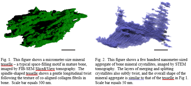
Structural hierarchy of bone – observed across multiple scales and in three dimensions – is essential to its mechanical performance. While the mineralized extracellular matrix of bone consists predominantly of carbonate-substituted hydroxyapatite, type I collagen fibrils, water, and noncollagenous organic constituents, it is largely the 3D arrangement of these inorganic and organic constituents at each length scale that endows bone with its exceptional mechanical properties. Based on recent volumetric imaging studies of bone using FIB-SEM tomography and STEM tomography, and earlier works spanning scales up to the level of whole bones, we illustrate the self-similarity of nested structural motifs across several orders of magnitude and link the hierarchical organization of bone to the ubiquity of spiral structure in Nature. We discuss the omnipresence of twisted, curved, sinusoidal, coiled, spiraling, and braided motifs in bone in at least nine of its twelve hierarchical levels – a visualization undertaking that has not been possible until recently with advances in 3D imaging technologies. We hypothesize that the twisting motif occurring across each hierarchical level of bone is linked to enhancement of function, rather than being simply an energetically favorable way to assemble mineralized matrix components. We propose that attentive, further consideration of twists in bone tissue and in the skeleton at different scales will likely enhance our understanding of structure–function relationships in bone.
- References
[1] Buss et al., Journal of Structural Biology X, Vol. 6, (2022), 100057.
[2] Reznikov et al., Science, Vol. 360, (2018), eaao2189.

